In this article
Author: Dr Károly Krkos
In our previous summary on IgG4 tests, we mentioned that antibodies of the IgG isotype are the antibodies found in the highest concentration in serum (they make up more than 70% of immunoglobulins)3. Of the four IgG subclasses, IgG1, IgG2 and IgG3 are able to form immune complexes with the antigen, activate complement (release pro-inflammatory cytokines) and bind strongly to FcγRI receptors (that is, enhance the antigen-clearing function of phagocytic cells).
IgG4, which represents the smallest fraction of IgG, differs from these in many of its properties: it is unable to take part in immune complex formation, activate complement or induce the release of inflammatory molecules4,20. This is because food sensitivity associated with IgG molecules is based on immune complex formation and the appearance of the phenomena characteristic of the associated inflammation (type III food sensitivity). To detect immune-mediated, food-induced hypersensitivity that causes complaints, food-specific antibodies must be sought in the IgG1, IgG2 and IgG3 subclasses23. This is why FOODTEST measures antibodies of all IgG classes produced against food components. By contrast, IgG4 formation cannot trigger an inflammatory reaction, and the IgG4 test is not suitable for identifying the foods that cause complaints (for setting up an elimination diet)9,23,26,27,28.
The properties of IgG4 have been studied intensively in recent years, and its role in physiological and pathological processes has come into the focus of interest.
The following characteristics have been demonstrated:
C1q-binding capacity
The spatial structure of the CH2 (heavy chain constant 2) domain, which plays a role in complement binding, differs in IgG4 from the same region in the other IgG subclasses2,32,38,39.
Figure 1: The difference between the parts of IgG1 and IgG4 involved in C1q binding (based on reference 38)
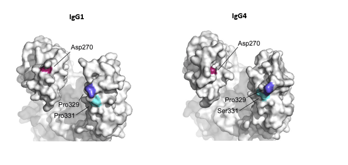
The asparagine at position 270 and the proline at position 329 are located elsewhere in space in IgG4 (compared with IgG1), while at position 331, which is in the same spatial position, IgG4 has serine instead of proline ® IgG4 is unable to bind the C1q component of the complement system (it cannot activate the classical pathway)
"Fab-arm" exchange
In the hinge region (CH2 domain) of IgG4, between the cysteines at positions 226 and 229 (which link the two half-molecules with inter-chain disulphide bridges), there is proline-serine instead of proline-proline (unlike in other IgG molecules). A further difference is that in the CH3 domain there is arginine at position 409 (instead of lysine). Because of these, the bond between the two chains is labile, so IgG4 easily splits into 2 half-molecules and recombines with another half-molecule of different specificity (differing in its variable region). The result is a functionally monovalent and bispecific IgG4, which is unsuitable for forming immune complexes25,32.
3. Fc-Fc interactions
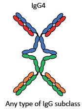
The arginine at position 409 also explains why the Fc region of IgG4 is able to bind to the Fc part of an IgG molecule of another class that is bound to a solid phase (e.g. a tissue structure). It presumably thereby helps to remove molecules of other IgG subclasses that damage the structures25.
Figure 3: Fc-Fc interaction (based on reference 25)
4. The affinity of IgG4 for FcγR receptors
Compared with molecules of the other IgG subclasses, it binds more weakly, as with FcγRI receptors, to FcγRIIa (histidine instead of arginine at position 131) and FcγRIIIa (valine replaces phenylalanine at position 158) receptors. It binds with greater affinity to the only inhibitory Fcγ receptor (FcγRIIb)5. It is completely unable to activate the FcγRIIIb receptor21.
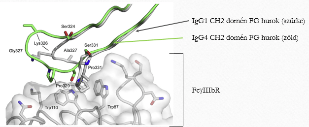
Figure 4: The inability of IgG4 to bind to the FcγRIIIb receptor can be explained by the difference between the CH2 domains of IgG1 and IgG4 (owing to the difference in the composition of the "FG loop" between positions 324 and 334) (based on reference 38)
a) The position of the proline at position 329 in the IgG1 FG loop allows it to wedge between tryptophans 87 and 117 in the receptor (a "proline sandwich"), whereas this is not true of the proline at position 329 in IgG4.
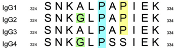
b) The amino acid sequence of the FG loop in molecules of the different IgG subclasses
In every IgG molecule other than IgG4 there is proline at position 331; in IgG4 there is serine here.
5. Affinity for the antigen
We already mentioned in our previous article ("On the IgG4 food intolerance test") that during the maturation of the immune response, the initial IgM production is followed by class switching to IgG3, then IgG1, in a prolonged immune response to IgG2 and finally to IgG4, while the affinity of the antibody for the antigen becomes ever stronger12,23,25, so IgG4 is able to bind its antigen with the greatest strength.
The role of IgG4 in various pathological conditions
A rise in IgG4 levels is a sign of immune tolerance during allergen immunotherapy (AIT)
Allergen immunotherapy (AIT) is used as a disease-modifying treatment for allergic conditions: the allergen is administered subcutaneously or under the tongue, first in small and then in increasing doses8,13. The method is used mainly in allergic rhinitis and rhinoconjunctivitis, allergic asthma and Hymenoptera venom allergy. The following changes can be observed as a result of the therapy:
- mast cell and basophil activity and degranulation decrease,
- changes in the isotype of anti-allergen antibodies: initially the IgE concentration rises and later falls, while the IgG4 concentration starts to rise immediately and this increase becomes continuous8,17,19,
- allergen-specific Treg and Breg cells are produced, while allergen-specific effector T cells and innate lymphocytes are suppressed,
- the number of tissue mast cells and eosinophils decreases (late effect).
During mast cell activation, the cell releases its granules containing histamine, enzymes (tryptase and chymase) and heparin into the surroundings. In addition, cytokines (TNF, IL4, IL-13) are released and lipid mediators (PGD2, LTB4, LTC4, LTD4, LTE4, PAF) are produced6.
Months after treatment, the early response to the allergen is significantly reduced, and the late response even more so.
Beekeepers who become resistant to bee stings after repeated stings have high levels of IgG4 produced against insect venom. (With few stings, the IgG1 level is raised.)
Allergen-specific IgG4
- competes with IgE on mast cells for binding the allergen; because of Fab exchange, IgG4 is bispecific, so the allergen cannot cross-link it8,15,
- IgG4 probably reduces the sensitivity of the antigen-presenting B cell, and thus of the T cell, and IgE-facilitated allergen presentation decreases [the activity of antigen-presenting B cells carrying the low-affinity IgE receptor (CD23) is enhanced by low levels of allergen in the presence of specific IgE; if IgG4 has been produced, a much larger amount of allergen is needed to activate allergen presentation]8,15,20.
- activates the blocking FcgRIIB receptors14,16,20,21
- does not bind complement and inhibits complement activation triggered by other IgG subclasses – anti-inflammatory effect8,25,32
- the IgE/IgG4 ratio is a good indicator for estimating the effectiveness of AIT8,22; a food-induced allergic reaction cannot be triggered if, alongside high IgE, the IgG4 concentration is higher22.
A rise in food-specific IgG4 levels can also be observed in eosinophilic oesophagitis (IgG4 against milk proteins, typically Bos d4, d5 and d8, possibly + IgG4 against wheat protein), which is why the role of high-pressure homogenisation in the disease becoming more common has been raised18,31. In the dietary treatment of the disease, a six- or seven-food elimination diet is used regardless of IgE or IgG4 positivity (see our previous article, "Mixed IgE/non-IgE allergy").
2. IgG4-related disease (IgG4-RD)
The main features of this group of diseases are: lymphoplasmacytic infiltration of the affected tissue with a predominance of IgG4-positive plasma cells, a degree of fibrosis, obliterative phlebitis and a raised eosinophil count10,29. The fibrosis shows a so-called "storiform" pattern (fibroblasts and inflammatory cells arranged like the spokes of a cartwheel). In many cases the serum IgG4 level is raised, but its absence does not rule out the disease.
In terms of pathogenesis, these are autoimmune conditions in which both B cells and T cells (mainly CD4+ and follicular T helper (Tfh) cells) play a role.
The disease is presumably triggered by several autoantigens. The most likely of these are:
- galectin-3,
- laminin 111,
- annexin A11.
Immune complex deposition is often seen in the affected organs. The largest numbers of cells observed in the affected tissues are CD4+ cytotoxic T lymphocytes (CD4+ CTL)11, which are maintained by continuous antigen presentation by B cells, and these cells presumably play a central role in the development of the condition. Through their IL-4 production, Tfh cells promote class switching to IgG4.
IgG4 appears to play no role in the pathogenesis. It is an immunoglobulin with an anti-inflammatory effect, and its increase is an accompanying phenomenon. A rise in IgG4 is not specific to IgG4-RD10,30; however, serum IgG4 concentrations above 2.8 g/l were associated with multi-organ involvement and an increased risk of relapse30,36.
The conditions belonging to this group of diseases were listed in our previous report ("On the IgG4 intolerance test").
3. IgG4-mediated autoimmune diseases
In some autoimmune diseases, the autoantibody produced belongs to the IgG4 subclass, and in these conditions it plays a causal role32. These conditions include:
- MuSK autoantibody-positive myasthenia gravis
- Pemphigus vulgaris and pemphigus foliaceus
Myasthenia gravis
Myasthenia gravis is a disease of the neuromuscular synapse: autoantibodies are produced against the acetylcholine receptors (AChR) needed for nerve-muscle signal transmission, resulting in progressive muscle weakness. There is also another form of the disease (occurring in about 40% of cases): these are AChR autoantibody-negative, MuSK autoantibody-positive conditions. MuSK is muscle-specific kinase. AChR formation and clustering require MuSK activation through the binding of agrin to Lrp4 (LDL receptor-related protein 4). MuSK autoantibodies can prevent this. While the disease based on AChR antibodies involves complement activation, MuSK autoantibodies are of the IgG4 type and do not activate complement. The treatment of the latter condition differs fundamentally from that of the AChR ab type, and it is more difficult to manage33.
Pemphigus vulgaris and pemphigus foliaceus
Pemphigus is an autoimmune blistering disease caused by autoantibodies against keratinocyte cell-surface proteins, desmoglein 3 (pemphigus vulgaris) and desmoglein 1 (pemphigus foliaceus), which belong to the IgG4 subclass34.
Other autoimmune conditions in which raised serum IgG4 concentrations are observed (in the late phase of the disease), but whose pathological role has not been proven: bullous pemphigoid and idiopathic membranous glomerulonephritis32.
4. The role of IgG4 in parasitic infections
IgE plays an important role in defence against parasites (helminths and some protozoa)7. When certain parasitic infections (schistosoma, roundworms, intestinal worms) become chronic, the parasites induce the host's immune system to respond in a protective way that dampens the immune defence: IgE production is replaced by IgG4 production, which acts in the direction of tolerance25,32.
5. The role of IgG4 in malignant diseases
In certain tumours (melanoma, extrahepatic cholangiocarcinoma, pancreatic, gastric and bowel cancer, glioblastoma), a rise in tumour-specific IgG4 levels is an unfavourable sign, as it indicates the development of tolerance towards the tumour25,32.
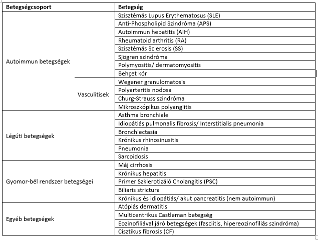
Other conditions associated with high serum IgG4 levels
Table (based on references 35 and 37)
Significantly higher total IgG4 levels were also found in people with autism spectrum disorder compared with the neurotypical group. The IgG4 level correlated with the degree of autism-specific impairment of social interaction24
Reduced serum IgG4 levels
IgG4 deficiency is common (it occurs in about 10% of the population3), but it is mostly asymptomatic. It may occur in isolation or combined with IgG2 or IgG2-IgA deficiency. Reduced IgG4 levels can be observed1 in:
- ataxia-telangiectasia,
- chronic mucocutaneous candidiasis,
- growth hormone deficiency,
- allergic colitis,
- Down syndrome.
References
- A P Knutsen: IgG subclass deficiency 2021, UpToDate, https://www.uptodate.com/contents/igg-subclass-deficiency/print?search=IgG&source=search_result&selectedTitle=1~150&usage_type=default&display_rank=1
- A M Davis, B J Sutton: Human IgG4: Structural perspective 2015, Immunological Reviews, 268:139-159 https://doi.org/10.1111/imr.12349
- R A Schwartz: Immunglobulin G Deficiency 2021, Medscape; https://emedicine.medscape.com/article/136897-print
- A P Knutsen, R Marsh: IgG subclasses: Physical properties, genetics, and biologic functions2022, UpToDate, https://www.uptodate.com/contents/igg-subclasses-physical-properties-genetics-and-biologic-functions/print?search=IgG%20subclasses:%20Physical%20properties,%20genetics,%20and%20biologic%20functions&source=search_result&selectedTitle=1~150&usage_type=default&display_rank=1
- M C Castells, L Bankova: Mast cells: Surface receptors and signal transduction 2021, UpToDate, https://www.uptodate.com/contents/mast-cells-surface-receptors-and-signal-transduction?search=IgG&source=search_result&selectedTitle=19~150&usage_type=default&display_rank=19
- S F Kemp: Pathophysiology of anaphylaxis 2022, UpToDate, https://www.uptodate.com/contents/pathophysiology-of-anaphylaxis/print?search=IgG&source=search_result&selectedTitle=14~150&usage_type=default&display_rank=14
- J Stokes, T B Casale: The biology of IgE 2020, UoToDate, https://www.uptodate.com/contents/the-biology-of-ige/print?search=IgG&topicRef=386&source=see_link
- M Akdis: Allergen immunotherapy for allergic disease: Therapeutic mechanisms 2020, UpToDate, https://www.uptodate.com/contents/allergen-immunotherapy-for-allergic-disease-therapeutic-mechanisms/print?search=IgG&source=search_result&selectedTitle=11~150&usage_type=default&display_rank=11
- J M Kelso: Unproven and disproven test for food allergy 2022, UpToDate, https://www.uptodate.com/contents/unproven-and-disproven-tests-for-food-allergy?search=IgG&source=search_result&selectedTitle=12~150&usage_type=default&display_rank=12
- H M Moutsopoulos, G E Fragoulis, J H Stone: Pathogenesis and clinical manifestation of IgG4-related diseases 2020, UpToDate; https://www.uptodate.com/contents/pathogenesis-and-clinical-manifestations-of-igg4-related-disease/print?search=IgG%20subclasses:%20Physical%20properties,%20genetics,%20and%20biologic%20functions&topicRef=3964&source=see_link
- Arata Takeuchi, Takashi Saito: CD4 CTL, a Cytotoxic Subset of CD4+T Cells, Their Differentiation and Function 2017, Frontiers in Immunology, 8:194, https://doi.org/103389/fimmu.2017.00194
- A M Collins, K J L Jackson: A temporal model of human IgE and IgG antibody function 2013, Frontiers in Immunology; https://doi.org/10.3389/fimmu.2013.00235
- M Larché, C A Akdis, R Valenta: Immunological mechanisms of allergen-specific immunotherapy 2006, Nature Reviews Immunology, 6:761-771; doi:10.1038/nri1934
- R T Strait, S C Morris, F D Finkelman: IgG-blocking antibodies inhibit IgE-mediated anaphylaxis in vivo through both antigen interception and FcIIB cross-linking 2006, J Clin Invest, 116(3):833-841; doi: 10.1172/JCI25575 During SIT, both allergen interception and the activation of the blocking FcRIIB receptors play a role in suppressing the allergic reaction.
- G L Piacentini, S Gueressi et al: A comparison between IgE and IgG4 (International Journal of Immunpathology and Pharmacology 2011, 24(4):1049-1056
- S Aschermann, A lux et al.: The other side of Immunglobulin G: suppressor of inflammation 2009, Clin Exp Immunol, 160:161-167; doi:10.1111/j.1365-2249.2009.04081.x
- S Piconi, D Trabattoni et al.: Effects of sublingual immunotherapy 2010, Journal of Immunology, 185:7723- 7730; doi: 10.4049/jimmunol.1002465
- T A E Platts-Mills, B Keshavarz et al.: An overview of relevance of IgG4 antibodies in allergic disease with a focus of food allergens 2021, Children, 8:418, doi: 10.3390/children8050418
- O Smoldovskaya, G Feyzkhanova et al.: Allergen-specific IgE and IgG4 patterns among patients with different allergic diseases 2018, World Allergy Organization Journal, https://doi.org/10.1186/s40413-018-0220-5
- W van de Veen, M Akdis: Role of IgG4 in IgE-mediated response 2016, American Academy of Allergy, Asthma and Immunology; http://www.elsevier.com/open-access/userlicense/1.0/
- L K James, S J Till: Potential Mechanisms for IgG4 Inhibition of Immediate Hypersensitivity reactons 2016, Curr Allergy Asthma Rep, 16:23; DOI 10.1007/s11882-016-0600-2
- G Noh, H-S Ahn et al.: The clinical significance of food-specific IgE/IgG4 in food-specific atopic dermatitis 2007, Pediatric Allergy and Immunology, 18:63-70; DOI: 10.1111/j.1399-3038.2006.00478.x
- C Meletis: The Clinical Utility of IgG4 testing 2019, US BioTek Laboratories 2019, https://www.usbiotek.com/blog/clinical-utility-igg4-testing
- A Enstrom, P Krakowiak et al: Increased IgG4 level in children with autism disorder 2009, Brain Behav Immun, 23(3):389-395; doi:10.1016/j.bbi.2008.12.005.
- R Bianchini, S N Karagiannis et al.: The Role of IgG4 in the Fine Tuning of Tolerance in IgE-mediated Allergy and Cancer 2020, International Journal of Molecular Science, 21:5017; doi:10.3390/ijms21145017
- S O Stapel, R Asero et al: Testing for IgG4 against food is not recommended as a diagnostic tool: EAACI task force report 2008, European Journal of Allergy and Clinical Immunology, https://doi.org/10.1111/j.1398-9995.2008.01705.x
- C Serrano-Moreno, N Brox-Torrecilla et al.: Diets for inflammatory bowel disease: What do we know so far? 2022, European Journal of Clinical Nutrition, 76:1222-1233, https://doi.org/10.1038/s41430-021-01051-9
- E Lavine: Blood testing for sensitivity, allergy or intolerance to food 2012, CMAJ; DOI: 10.1503/cmaj.110026
- K-H Yu, T-M Chan et al.: Diagnostic Performance of Serum IgG4 levels in Patients with IgG4-related disorders 2015, Medicine, 94(41); DOI: 10.1097/MD.0000000000001707
- E L Culver, R Sadler et al: Elevated Serum IgG4 levels in Diagnosis, Treatment Response, Organ Involvement, and Relapse in a Prospective IgG4-Realted Disease UK Cohort 2016, The American Journal of Gastroenterology; doi: 10.1038/ajg.2016.40
- E C McGowan, T A E Platts-Mills, J M Wilson: Food Allergy, Eosinophylic Esophagitis and the Enigma of IgG4 2020, Ann Allergy, Asthma Immunol, 122(6):563-564; doi:10.1016/j.anai.2019.03.024
- D C Trampert, L M Hubers et al: On the role of IgG4 in inflammatory conditions: Lessons for IgG4-related disease 2017, BBA – Molecular Basis of Disease; http://dx.doi.org/10.1016/j.bbadis.2017.07.038
- M G Hulibers, W Zang et al.: MuSK IgG4 autoantibodies cause myasthenia gravis by inhibiting binding between MuSK and Lrp4 2013, PNAS, 110(51):20783-20788, https://doi.org/10.1073/pnas.1313944110
- Yuko Futei, Masayuki Amagai et al.: Predominant IgG4 subclass autoantibodies in pemphigus vulgaris and foliaceus 2001, Journal of Dermatological Science, 26:55-61, https://doi.org/10.1016/S0923-1811(00)00158-4
- M Ebbo, A Grados et al: Pathologies Associated with Serum IgG4 Elevation 2012, International Journal of Rheumatology, Article ID:6028098; doi:10.1155/2012/602809
- I Mizushima, M Konishi et al: Serum IgG4 levels at diagnosis can predict unfavorable outcomes of untreated patients with IgG4 disease 2021, Scientific Reports, 11:13341; https://doi.org/10.1038/s41598-021-92814-8
- J H Ryu, R Horie et al: Spectrum of Disorders Associated with Elevated Serum IgG4 Levels Encountered in Clinical Practice 2012, International Journal of Rheumatology, Article ID:232960; doi:10.1155/2012/232960
- A M Davies, T Rispens et al.: Structural Determinants of Unique Properties of Human IgG4 Fc 2013, Journal of Molecular Biology, http://dx.doi.org/10.1016/j.jmb.2013.10.039
- A M Davies, R Jefferis, B J Sutton: Crystal structure of deglycosylated human IgG4-Fc 2014, Molecular Immunology, 62:46-53, https://doi.org/10.1016/j.molimm.2014.05.05
SIBO and food intolerances: is food necessarily to blame?
Bloating after milk, bread or onions? The way the digestive system works may also play a role behind these symptoms. Dr Anikó Hovanecz’s article expla...
A corporate health programme in practice – the SFC CEO’s experience
What does caring for employees and their family members mean for a company leader? Through his personal experience, Gábor Horváth, CEO of SFC, explain...

Don't just get the school bag ready
Starting school is not only about new exercise books and a changed daily routine but also about reorganising family meals. The FOODTEST Children's pac...