In this article
Author: Dr Károly Krkos
The role of mast cells has been studied most intensively in atopic reactions and parasitic infections. More recent studies also suggest their involvement in autoimmune inflammatory processes, atherosclerosis and resistance to bacterial infections3.
Allergens act through the cross-linking of IgE molecules bound to the FcεRI receptor. The signalling that develops during activation requires two members of the Src family of kinases: Lyn and Fyn. Fyn activates STAT5, which is needed for mast cell survival and cytokine production. The regulatory cytokines IL-10 and TGFβ can inhibit Fyn and STAT5 production – presumably through the induction of specific miRNAs3.
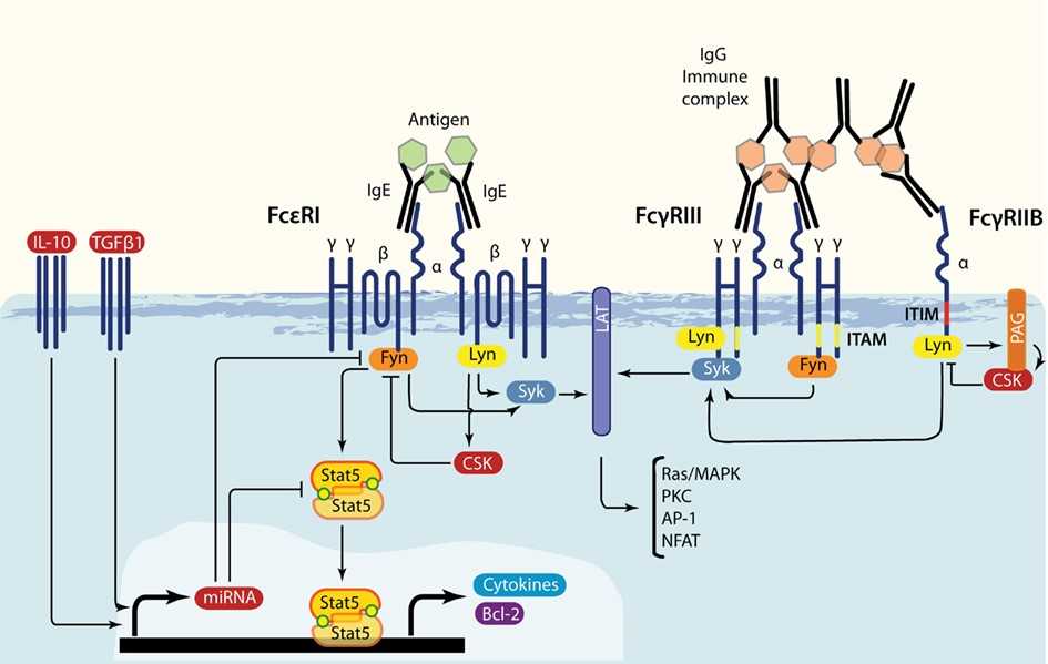
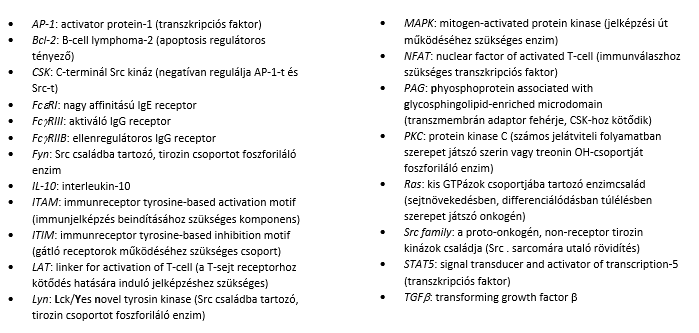
Mast cells also express FcγRI, FcγRIII and FcγRIIB receptors on their surface. The FcϒRI and FcγRIII receptors bind host-directed IgG or IgG–protein immune complexes and, through signalling similar to that induced via the FcεRI receptor, trigger the production of pro-inflammatory molecules. The FcγRIIB receptor mediates a counter-regulatory effect, the production of anti-inflammatory molecules3 (see Figure 1).
Figure 1 Signalling through the activation of the FcεRI, FcγRIII and FcγRIIB receptors3
The maturation of mast cells is governed by the tissue microenvironment and by cytokines (mainly stem cell factor, SCF). Other molecules involved in mast cell maturation are NGF (nerve growth factor) and neurotrophin-3. Molecules that direct mast cells to the site of the process (chemoattractants) include SCF, MCP-1 (monocyte chemoattractant protein-1, or CCL2) and RANTES (Regulated upon Activation, Normal T-cell Expressed and Secreted, or CCL5)1.
Activated mast cells store vasoactive and pro-inflammatory substances in their granules: histamine, serotonin, tumour necrosis factor (TNF), kinins and proteases. The proteases cleave histamine-releasing peptides (histamine release peptides, HRP) from plasma albumin and also activate the protease-activated receptor (PAR), which increases permeability1.
During activation the following molecules are also synthesised: leukotrienes, prostaglandins and platelet-activating factor (PAF). In addition, a few hours after stimulation numerous cytokines (IL-1, -2, -3, -5, -6, -8, -9, -13, TNF) and VEGF (vascular endothelial growth factor) are produced1.
Mast cells can not only release the substances stored in their granules but can also secrete bioactive substances (IL-6, eicosanoids, VEGF, TNF) selectively without degranulation, which plays a role in the following conditions: arthritis, atopic dermatitis and multiple sclerosis1.
CRH, the hypothalamic hormone that activates the pituitary gland's production of ACTH (adrenocorticotropic hormone) and the adrenal cortex, is also produced in immune cells and mast cells1.
Mast cells play a role in innate and acquired immunity as well as in autoimmunity. They contribute to the maturation of Th17 cells, which play a key role in autoimmunity1.
Figure 2: The role of mast cells in various inflammatory conditions1:
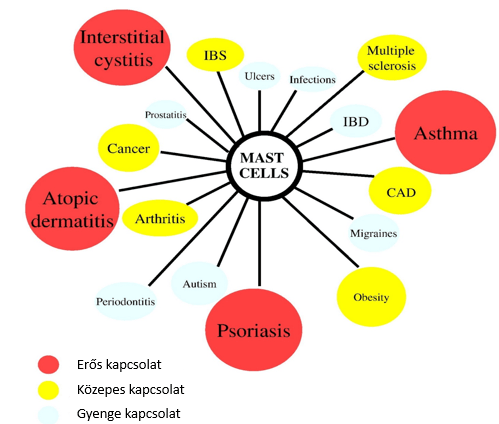
CAD, coronary artery disease; IBD, inflammatory bowel disease; IBS, irritable bowel syndrome
The role of mast cells in functional gastrointestinal (GI) disorders (FGIDs)
In the intestines of people with functional gastrointestinal disorders (FGIDs), mainly mast cell and eosinophil infiltration can be observed beneath the mucosa, and there are more lymphocytes between the absorptive epithelial cells than in healthy people (intraepithelial lymphocytes). Disruption of the balance of gut–brain communication leads to motility disorders and hypersensitivity. FGIDs are diagnosed on the basis of symptoms and assessed according to the Rome criteria. The two most common conditions in this group are functional dyspepsia (FD) and irritable bowel syndrome (IBS), which affects around 16–26% of the world's population2. They are characterised by low-grade mucosal inflammation associated with impaired intestinal permeability, and by neuronal sensitivity2.
In the GI tract, mast cells play a role in:
- regulating vascular and epithelial permeability,
- ion secretion,
- angiogenesis,
- intestinal motility,
- the development of fibrosis and tissue repair,
- innate and acquired immunity,
- cell recruitment,
- defence against bacteria,
- pain perception.
Regulation of the activation of mast cells (mastocytes, MCs)
The most potent stimulus for mast cells is when an antigen cross-links the IgE molecules bound to the FcεRI receptors on their surface. This results in degranulation and cytokine production.
Mast cells can also bind IgG molecules (FcγRI), light chain molecules (IgLC) and complement products, and they also carry TLRs (toll-like receptors).
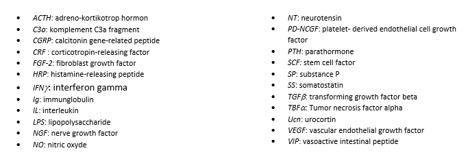
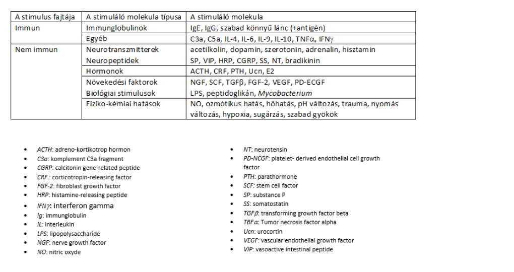
Table 1: Molecules that activate mast cells: neurotransmitters, neuropeptides, growth factors and hormones:
Factors and mechanisms causing MC activation in the gut
Food antigens triggering MC activation
28% of patients with FGIDs report a worsening of symptoms within 15 minutes of eating, and 93% within 3 hours of eating. Although atopy is more common in some IBS patients, food allergy shows no convincing association with FGIDs. Food intolerance may also develop as a result of IgG-mediated sensitisation of mast cells, but academic medicine does not yet consider the role of IgG-mediated immune reactions to be proven.
When the suspected food is applied endoscopically directly onto the duodenal mucosa of IBS patients, damage to the absorptive epithelium can be observed immediately, and the intervillous space and the number of intraepithelial lymphocytes increase. Withdrawing these foods leads to symptom relief within 1 year in 74% of patients. At the same time, the response to food is partly regulated by neuroendocrine factors (such as the serotonergic response). Mast cells can synthesise and secrete serotonin from tryptophan, and serotonin is also one of the chemotactic factors for MCs. Foods high in serotonin (cheeses, meats, soya, cereals, nuts, vegetables) can cause unpleasant effects. Spicy foods can provoke IBS. Capsaicin is a ligand of the TRPV1 (transient receptor potential vanilloid 1) receptors on nociceptive afferent C fibres. A higher density of sensory fibres expressing TRPV1 receptors has been detected in FGID patients (compared with healthy people). The proximity of mast cells to sensory nerve fibres expressing TRPV1 receptors, together with the ability of capsaicin to modulate mast cells, may explain the pain that develops after eating spicy foods. At the same time, desensitisation can be achieved with a diet high in capsaicin: bloating and pain decrease after a 6-week course.
The effect of infections
Infectious gastroenteritis is associated with an increased risk of developing FD and IBS, but it is not known what factors cause the complaints to become chronic. Mast cells take part in both innate and acquired immune processes against bacteria, viruses, parasites and fungi. In patients with post-infectious, diarrhoea-predominant IBS (IBS-D), the proximity of MCs and plasma cells points to immunoglobulin-induced activation of the mast cells.
The role of stress
Chronic stress leads to mast cell activation. Stress mediators such as CRF and related peptides alter ion and water secretion as well as the paracellular and transcellular permeability of the colon – among other things through nerve–mast cell interaction.
Table 2: Conditions that alter – mainly increase – the number of mast cells in the GI tract
- Allergic diseases: chronic urticaria, food allergy, atopy, hereditary angioedema
- Mastocytosis and mast cell activation syndrome
- Coeliac disease: mast cell infiltration is increased in the early phase and decreased at a later stage
- Tumours: neuroendocrine tumours, lymphoma, epithelial cancers, carcinoid syndrome
- Infectious gastrointestinal inflammation: Helicobacter pylori gastritis, infectious gastritis, parasitic infections
- IBS, lymphocytic colitis
- Intestinal pseudo-obstruction, diverticulitis
- Vasculitis, amyloidosis, medicines
The pathophysiology of FGIDs is characterised primarily by increased activity of mast cells (rather than an increased number of them).
In the human gut, mast cells are located close to sensory nerve endings rich in neuropeptides and TRPV1 receptors. Afferent innervation of intestinal mast cells can trigger the release of histamine and proteases, and these mediators increase the sensitivity of spinal afferent terminals in a paracrine manner.
Activation of the afferent nerve can trigger myenteric cholinergic depolarisation, which in turn alters motor function.
Figure 3: Mast cell–nerve interaction. The bidirectional interaction between mast cells and nerves modifies peristalsis and the pain signal. The bioactive, pro-inflammatory substances released from the mast cell have various effects on the nerve ending (activation, sensitisation, recruitment of nociceptors to the cell membrane, neurogenic inflammation), which ultimately lead to visceral hypersensitivity. Neuronal activation triggers the release of neuropeptides and neurotransmitters, which in turn further activate the mast cell.
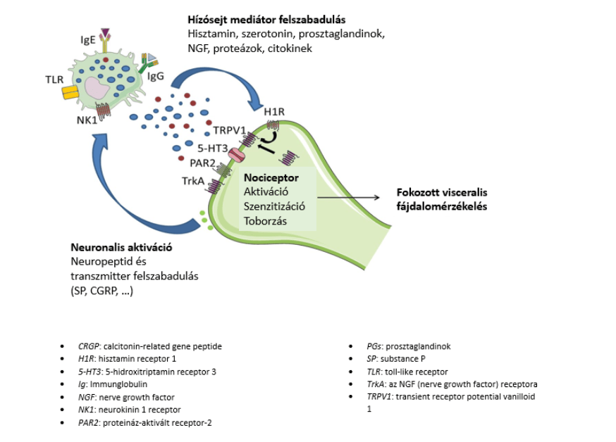
In IBS patients, the level of mast cell-derived mediators is increased in the intestinal mucosa and submucosa, which leads to activation of afferent nerves, increased visceral pain perception and altered intestinal motility (diarrhoea or constipation).
The role of mast cells in regulating intestinal barrier function
The role of mast cells in changes in intestinal permeability was first suggested when the administration of a mast cell stabiliser reduced the increased transport of ions and macromolecules across the intestinal epithelial cells.
The mechanism of the mast cell effect is not fully understood, but the ability of substances released from mast cells to break down TJ proteins may play a role. In IBS patients, the expression of several tight junction (TJ) proteins was reduced compared with the control group; in IBS-D patients this was associated with mast cell activation and clinical symptoms. Tryptase released from MCs activates the PAR2 receptor, which leads to changes in the TJs and thus increases the permeability of the gut to macromolecules. Other mast cell mediators – histamine, chymase and prostaglandin D2 – influence chloride ion and water secretion and the permeability of the intestinal epithelium (Figure 4).
Figure 4: Interaction between mast cells and the components of intestinal barrier function: TJ modulation and secretory response, recruitment of other immune cells, altered vascular function (permeability, blood flow), peristalsis and pain signal.
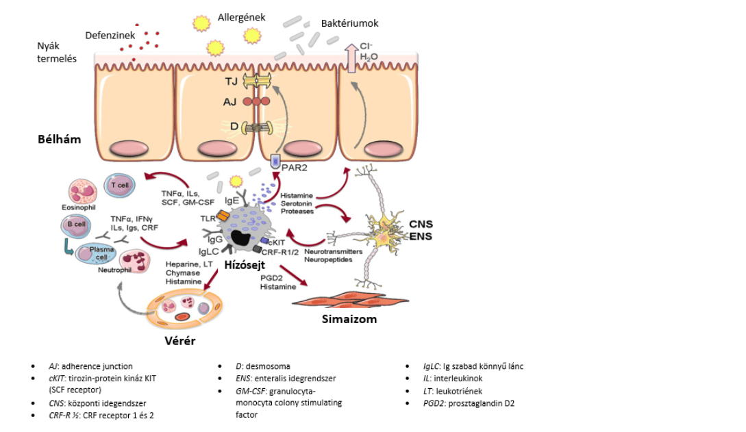
The interaction between mast cells and the gut–brain network plays a role in the development of symptoms in some FGID patients.
Treatments targeting mast cells in FGID patients
The mast cell stabiliser disodium cromoglycate (DSCG) abolishes the increase in small intestinal permeability associated with acute psychological stress.
Anti-inflammatory mesalazine reduces the number of MCs by 36% and significantly reduces the number of total immune cells and T cells in the colonic mucosa.
Palmitoylethanolamide and other cannabinoid receptor inhibitors are effective in controlling pain, motor disorders and inflammation by modulating neuronal and non-neuronal cells (including mast cells).
750 mg/day of slow-release vitamin C increases histamine breakdown and inhibits mast cell degranulation.
Natural flavonoids (fisetin, kaempferol, quercetin, rutin, luteolin) and berberine (a benzylisoquinoline alkaloid) inhibit the release of mediators from mast cells (protecting the epithelial barrier).
References
- T C Theoharides, K-D Alysandratos et al.: Mast cells and inflammation, Biochim Biophys Acta 2012, 1822(1):21-33, https://doi.org/10.1016/j.bbadis.2010.12.014
- N A Pullen, Y T Falanga et al.: The Fyn-STAT5 pathway: a new Frontier in IgE- and IgG-mediated mast cell signalling, Frontiers in Immunology 2012, 3: Article 117, https://doi.org/10.3389/fimmu.2012.00117
- M M Wouters, M Vicaro, J Santos: The Role of Mast cells in functional GI Disorders, Gut 2016, 65:155-168 http://dx.doi.org/10.1136/gutjnl-2015-309151
SIBO and food intolerances: is food necessarily to blame?
Bloating after milk, bread or onions? The way the digestive system works may also play a role behind these symptoms. Dr Anikó Hovanecz’s article expla...
A corporate health programme in practice – the SFC CEO’s experience
What does caring for employees and their family members mean for a company leader? Through his personal experience, Gábor Horváth, CEO of SFC, explain...

Don't just get the school bag ready
Starting school is not only about new exercise books and a changed daily routine but also about reorganising family meals. The FOODTEST Children's pac...