In this article
Author: Dr Károly Krkos
In our previous summary we already mentioned the classification of problems that fall under food sensitivity (Figure 1) and one of the immune-mediated disease groups (non-IgE allergies). In this article we would like to briefly describe conditions in which IgE production is increased but other mechanisms dominate the pathogenesis of the disease group (“mixed IgE/non-IgE allergy”).
Figure: Classification of food sensitivities
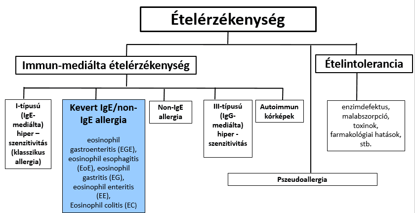
In diagnosing mixed IgE/non-IgE and non-IgE allergies, the primary role is played by taking a thorough, detailed history, which in many cases needs to be supplemented by imaging, endoscopic methods and examination of biopsy samples. This group includes the eosinophilic gastrointestinal diseases (EGID). Depending on the location of the disease, eosinophilic oesophagitis (EoE), eosinophilic gastritis (EG), eosinophilic gastroenteritis (EGE), eosinophilic enteritis (EE) and eosinophilic colitis (EC) are distinguished1-3,8.
EGID is considered a rare disease; its prevalence has been estimated at 22–28 per 100,000 people (US)5, but based on insurance data it appears to be even lower:
- EG: 6.3/100,000 (in children over 5 years of age)
- EGE: 8.4/100,000 (more common in children under 5)
- EC 3.3/100,000
In adults it can occur at any age, but the peak frequency is observed in the 30–50 age group1-3,5.
Eosinophilic gastroenteritis (EGE)
Its pathogenesis is poorly understood. In 50–70% of patients the condition is associated with atopic diseases1-3, and in EG and EGE raised food-specific serum IgE levels are common. Although in EG/EGE this IgE level does not clearly indicate the triggering food, an elemental or elimination diet is nevertheless effective and improves the clinical, endoscopic and histological picture. In people with such a disease, food triggers the activation of IL-5-producing, allergen-specific T helper 2 (Th2) cells, which leads to the accumulation of eosinophils in the gut. The eotaxins, chemokines responsible for recruiting eosinophils, play a role in this. The persistence of eosinophils is maintained by the interleukins IL-3 and IL-5 and by granulocyte-macrophage colony-stimulating factor (GM-CSF). The presence of eosinophils leads to local inflammation, because cytotoxic cations (e.g. major basic protein, MBP) are released from them. The synthesis of Th2 cytokines (IL-4, IL-5, IL-13) and of the eosinophil chemokine eotaxin-3 increases1.
In terms of its clinical forms, it can affect the gastrointestinal tract from the oesophagus to the colon. Eosinophilic gastritis (EG) is localised to the antrum and body of the stomach. Eosinophilic gastroenteritis (EGE) typically affects the distal antrum and the proximal part of the small intestine. 50–70% of EG/EGE patients have some atopic disease (asthma, eczema, hay fever). The symptoms depend not only on the location of the disease but also on which layer of the gut wall is infiltrated by eosinophils. When the mucosa is involved, the leading symptoms are abdominal pain, nausea, vomiting, early satiety and diarrhoea. Weight loss (³ 2.4 kg) is seen in one third of patients. Diffuse small-intestinal involvement is associated with malabsorption and protein-losing enteropathy. Disease of the muscle (muscularis) layer results in thickening of the bowel wall and impaired motility, with consequent bowel obstruction, nausea, vomiting and bloating. Eosinophilic inflammation of the oesophagus can lead to stricture, difficulty swallowing and regurgitation of undigested food (pseudoachalasia). Obstruction or perforation of the gastric outlet, the small intestine and, rarely, the colon may occur. Disease of the tissue beneath the outer serous membrane (subserosa) is associated either with the appearance of fluid in the abdominal cavity (ascites) alone, or with symptoms typical of mucosal and muscularis disease as well. Eosinophils can be seen in the ascitic fluid.
Table: Clinical forms of EGE (based on A A Rached, W El Haj: Eosinophilic gastroenteritis: Approach to diagnosis and management – reference 2)
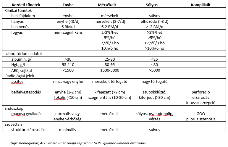
Laboratory abnormalities: in about 80% of patients the relative and absolute eosinophil counts are raised (5–35% and an average of 1,000 eosinophils/ml respectively). The mucosal and subserosal types are associated with higher eosinophil counts than the muscularis type1,5. In mucosal EG/EGE, carbohydrate malabsorption (abnormal D-xylose test), increased fat excretion (raised faecal fat content) and prolonged prothrombin time can be observed. Increased mucosal permeability can lead to protein-losing enteropathy, with low serum albumin levels. Anaemia is related to impaired iron absorption and occult gastrointestinal bleeding (iron deficiency). A moderately raised erythrocyte sedimentation rate is observed in about a quarter of patients. Serum IgE levels may be raised (mainly in childhood cases).
Endoscopy may show mucosal ulceration, polyps and pseudopolyps, sometimes signs of inflammation, villous atrophy, submucosal oedema and fibrosis. Because the process is patchy, samples must be taken from at least 5–6 sites, from both normal and abnormal areas, for histological examination3. Full-thickness samples (in non-mucosal disease) can be obtained by surgical exploration.
Evaluation of the histological picture is fundamental to the diagnosis. The most important diagnostic criterion is eosinophilic infiltration of the tissues, with other diseases causing eosinophilic infiltration ruled out. It is important that only the gastrointestinal tract is involved1,3. With the exception of the oesophagus, the entire gastrointestinal tract contains eosinophils, and the eosinophil counts characteristic of healthy people in the individual segments have not yet been clearly established. Microscopic evaluation must always be performed by a pathologist with sufficient experience in this field. M H Collins established the following abnormal eosinophil count values4:
- stomach: ≥30 eosinophils/high-power (400x) field (HPF)
- duodenum: ≥ 30 eosinophils /HPF
- ileum: >56 eosinophils/HPF
- ascending colon: >100 eosinophils /HPF
- transverse and descending colon: >84 eosinophils /HPF
- sigmoid colon and rectum: >64 eosinophils /HPF
Imaging shows thickening of the gastric and/or bowel wall and irregular narrowing of the bowel lumen (mainly in the distal antrum and the proximal small intestine). These are not specific to the disease1,3. CT shows oedema of the small intestine and the ileocaecal segment, which becomes visible as the characteristic “halo” and “araneid limb-like” signs18 (the images are based on the article by Xing Huang, Xiangqi Xiao et al: Halo sign and araneid limb sign in eosinophilic enteritis 2020, Lancet Gastroenterology and Hepatology):
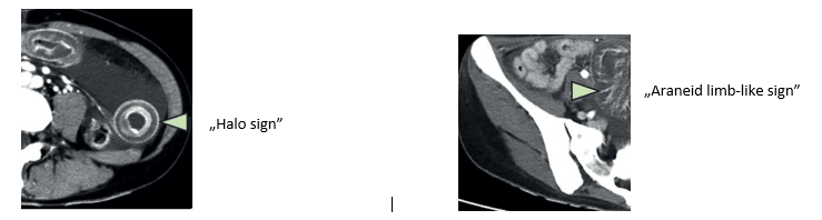
Abdominal ultrasound and CT can detect any fluid in the abdominal cavity (ascites), which is sometimes associated with localised lymph node enlargement. These signs can also be seen in Crohn’s disease, lymphoma and granulomatous diseases1,3. The “halo” and “araneid limb-like” signs are characteristic of inflammation (and help to distinguish the disease from neoplastic processes)3. SPECT/CT imaging with 99mTc-HMPAO-labelled white blood cells plays a role in assessing the extent of the disease and in monitoring the response to treatment3.
The diagnostic procedures mentioned above must be supplemented so that conditions with similar symptoms can be distinguished (differential diagnosis):
Drug-induced changes: acetylsalicylic acid (ASA), sulphonamides, penicillin, cephalosporins, carbamazepine, azathioprine, L-tryptophan and gold salts can cause similar tissue (and peripheral) eosinophilia5.
Intestinal infections: parasitic infections: Ancylostoma, Anisakis, Ascaris, Strongyloides, Toxocara, Trichiuris, Capillaria, Trichinella; fungal infection: basidiobolomycosis can be identified from stool or by serological testing. The clinical picture of Ancylostoma caninum infection mimics EGID most closely (eosinophilic infiltration of the bowel wall and ascites develop)1. Charcot-Leyden crystals derived from eosinophil granules can also be found in the stool.
Malignant diseases: lymphoma, gastric cancer and colon cancer may be accompanied by peripheral eosinophilia, the disease may cause obstruction, and imaging may show thickening of the bowel wall. These diseases can be distinguished by endoscopy and histological examination of full-thickness biopsy samples.
Inflammatory bowel diseases (IBD): Crohn’s disease and ulcerative colitis may sometimes be accompanied by eosinophilia and eosinophilic infiltration of gastrointestinal tissues. Endoscopy may show characteristic architectural distortion, focal neutrophilic exudates and granulomas.
Hypereosinophilic syndrome (HES): an idiopathic condition characterised by marked eosinophilia (<1500 eosinophils/ml for more than 6 months). It is rarely dominated by gastrointestinal complaints. In HES, however (unlike in EGID), several organs (heart, lungs, brain, kidneys) are involved. If HES is suspected, the help of a haematologist or allergist should be sought to rule out the disease. Polyarteritis nodosa (PAN): another condition with eosinophilia and abdominal pain, which is systemic (unlike EGID), with a markedly raised erythrocyte sedimentation rate and perivascular eosinophil accumulation in the biopsy sample.
Eosinophilic granulomatosis with polyangiitis (Churg-Strauss syndrome): EGID with abdominal pain, diarrhoea and gastrointestinal bleeding may appear before or at the same time as Churg-Strauss syndrome. The cardinal symptom of eosinophilic granulomatosis with polyangiitis is asthma, which precedes the onset of the vascular phase by 8–10 years.
Eosinophilic granuloma (Langerhans cell histiocytosis, LCH; old names no longer in use: histiocytosis X, Letterer-Siwe disease, Hand-Schüller-Christian disease): LCH is a neoplastic histiocytic disease that most often affects the bones and skin, but can also appear in the bone marrow, spleen, liver, lungs, pituitary gland, central nervous system and other organs. It is seen mainly in early childhood. The granulomatous appearance of the biopsy sample distinguishes it from EGID.
Its treatment has not yet been clearly established and is significantly influenced by the severity of symptoms and the presence or absence of malabsorption.
Dietary intervention is based on an empirical elimination diet: the six-food (soya, wheat, egg, milk, nuts and oilseeds, fish/seafood)1,3 or seven-food (the former + red meats)3 elimination diet, and in more severe cases an elemental diet (with calcium and vitamin D supplementation)1,3. With the latter, about ¾ of patients go into remission, but even this does not guarantee that symptoms will not return33. In disease with mild symptoms, targeted elimination based on specific IgE testing may also be tried3, but apart from mild cases no allergy test helps in putting together an effective diet1. The effectiveness of dietary treatment has been observed most clearly in mucosal-type EGE31; muscularis and subserosal disease are less dependent on food allergy32. The effectiveness of dietary treatment is indicated if the number of eosinophils in the blood falls by at least half within 4–6 weeks5.
Medication is necessary. Steroids come first. As initial therapy, prednisolone at a dose of 30–40 mg a day for 6–8 weeks results in remission in more than 90% of cases; however, gradually reducing the dose often leads to symptoms returning, in which case maintenance treatment of 1–10 mg a day is set up, or prednisolone is replaced with budesonide. The advantage of the latter is that, given as a slow-release enteric capsule, it causes fewer side effects. Treatment may be supplemented with a leukotriene receptor antagonist also used in treating asthma (montelukast sodium), a mast cell stabiliser [sodium cromoglycate (cromolyn, SCG)] and a second-generation antihistamine (ketotifen), immunomodulators (azathioprine, 6-mercaptopurine) and proton pump inhibitors. In recent years, new biological treatments (reslizumab – IL-5 antibody, infliximab – TNFα antibody, omalizumab – anti-IgE, suplatast tosilate – selective Th2 cytokine inhibitor), intravenous immunoglobulin, α-interferon (IFNα) and faecal microbiota transplantation have also been tried3.
Surgical treatment must be used in cases of perforation, intussusception or bowel obstruction.
Figure: Diagnostic algorithm for EGE (based on A A Rached, W El Haj: Eosinophilic gastroenteritis: Approach to diagnosis and management – reference 2)
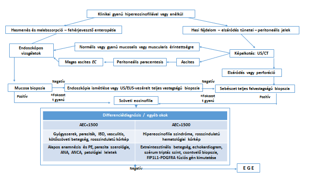
AEC: absolute eosinophil count; EC: eosinophil count; US: ultrasound; PE: physical examination
The course of the disease is chronic and fluctuating, but its prognosis is good, and malignant tumours do not develop on its basis1,3.
Eosinophilic oesophagitis (EoE)
The disease is localised to the oesophagus. Its symptoms resemble gastro-oesophageal reflux disease, but the condition does not improve with the usual (medical and surgical) treatment for reflux6-8. Diagnosis is based on the clinical picture together with examination of an oesophageal biopsy sample. More than 15 eosinophils are seen per high-power field4,6-8, and other causes of eosinophilia can be ruled out6-8.
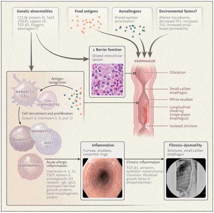
Its pathogenesis involves environmental factors (birth by caesarean section, premature birth, antibiotic treatment in infancy, food allergy, lack of breastfeeding, atopic diseases, altered microbiome), genetic predisposition (the genes for thymic stromal lymphopoietin – TSLP, eotaxin-3 and calpain-14), impaired barrier function (underproduction of filaggrin, zonulin-1 and desmoglein-1), deterioration of barrier function (enlarged intercellular spaces), increased Th2 activity, and recruitment of eosinophils, basophils, mast cells and NK cells. IgG4 produced against the four most common allergenic foods can be observed in the oesophageal epithelium7. IgE testing does not help in uncovering the triggering foods6-8. Concentric rings, furrows, and dilated and narrowed segments appear in the oesophagus, which can be linked to fibrosis developing on the basis of chronic inflammation (see Figure 2)
Figure: Pathology of eosinophilic oesophagitis (based on G T Furuta: Eosinophilic Esophagitis – reference 7)
In childhood the clinical picture is characterised by feeding difficulties, nausea, vomiting, abdominal pain and faltering growth. In teenagers and adults the leading symptoms are difficulty swallowing, food getting stuck, chest pain that does not improve with antacids, and chronic reflux6-8. Rarely, the oesophagus may even rupture from forceful retching (Boerhaave syndrome).
Three subtypes have been described6:
- EoEe1 – mild subgroup: normal-appearing oesophagus with mild histological, endoscopic and molecular changes
- EoEe2 – inflammatory endotype: increased expression of inflammatory cytokines, with steroid-responsive and steroid-refractory phenotypes
- EoE3 – fibrostenotic endotype: narrow-calibre oesophagus with severe abnormal endoscopic findings and histological changes, and reduced epithelial differentiation genes.
Endoscopy shows white exudates, mucosal oedema, linear furrows, oesophageal rings and strictures. Because of chronic remodelling, a “crêpe-paper oesophagus” develops. A validated endoscopic scoring system has been developed for evaluation (edema, rings, exudates, furrows and strictures scores – EREFS). It is advisable to take biopsy samples from at least 4–5 sites during the examination6-8.
A barium contrast X-ray is the most suitable method for assessing strictures, which are often not detected at endoscopy.
Histological examination is also fundamental to the diagnosis of EoE. Besides tissue eosinophilia (>15 eosinophils/high-power field), its features are mild to moderate basal cell hyperplasia, elongation of the vascular papillae, dilated intercellular spaces and intraepithelial lymphocytosis.
Dietary and medical treatment, similarly to that described in the EGE section, consists of an elemental or 6-food elimination diet, or steroids (prednisolone, budesonide)6-8. More recently, biological therapies have also been tried8. Proton pump inhibitors are also recommended; although in many cases reflux symptoms do not resolve with them, they reduce cytokine production (in addition to reducing gastric acid secretion). Cases of eosinophilic oesophagitis that respond to proton pump inhibitors are regarded as a proton pump inhibitor-responsive subtype. In the case of significant stricture, the above is usually supplemented by oesophageal dilation7.
Eosinophilic gastritis (EG)
The leading symptoms of the form of EGID localised to the stomach are epigastric pain, nausea, vomiting and weight loss. Anaemia and protein-losing enteropathy may also develop9-11. Usually the fundus and the antrum are affected (so samples need to be taken from both areas). Endoscopy shows erythema and ulceration, and the biopsy sample shows tissue eosinophilia. Of the other pathological conditions to be ruled out (parasitic infections, haematological and lymph node disorders, IBD), Helicobacter pylori infection and Crohn’s disease are the most common. In a study of biopsy findings, the histological results of 60 EG patients and 135 control patients were compared. In the healthy group, the number of eosinophils per 1 mm2 was <55 in all but 1 patient, whereas this value ranged between 127 and 2108 in the EG group10 (this corresponds to 30–500 eosinophils per high-power field, since its area is 0.237 mm2)9. Given that only 2 patients in the patient group had values <190 (one 127, the other 168 eosinophils/mm2), a value of ≥190/mm2 can be considered the lower limit for EG. The eosinophil density of patients with Crohn’s disease and those with Helicobacter pylori infection differed only minimally from that of the healthy population: the maximum was 203/mm2 (H. pylori) and 219/mm2 (Crohn’s disease)10. Peripheral eosinophilia occurs in 23–86% of cases; its absence does not rule out the condition, and its degree is not related to disease activity9,10. Its treatment is the same as described in the EGE section9-11.
Eosinophilic enteritis (EE)
The form of EGID localised to the small intestine (EE) is a very rare condition (its prevalence is thought to be 1/100,000). It is more common in men, and its peak age of onset is put between 30 and 40 years. The complaints are non-specific, and about 2 years pass from their appearance to diagnosis12. Presenting symptoms: abdominal pain, nausea/vomiting, diarrhoea, ascites, but jaundice and gastrointestinal bleeding may also develop. Complications requiring surgery (obstruction, stricture, peritonitis) may also occur. When the mucosa is involved, abdominal pain, diarrhoea and bleeding are typical; with disease of the muscularis layer, symptoms of obstruction are typical; subserosal infiltration is characterised by ascitic fluid rich in eosinophils, distension and abdominal pain. Frequency of the subtypes32: mucosal: 57.5%; muscularis: 30%, subserosal: 12.5%.
When the proximal small intestine is involved, protein-losing enteropathy (PLE) develops with hypoalbuminaemia, malabsorption (fatty stools, trace element and vitamin deficiency) and iron-deficiency anaemia. Peripheral eosinophilia (>500 eosinophils/ml) has been observed in 70–80% of cases, reaching the highest levels in the subserosal form. Serum IgE is high in about half of patients. Diagnosis requires upper gastrointestinal endoscopy and ileocolonoscopy as well as biopsy, supplemented by ultrasound, CT and MRI imaging. A contrast X-ray of the small intestine clearly shows wall thickening, stricture and dilatation, as well as ulcerative inflammation of the terminal ileum. In addition, the conditions mentioned above, which may present with similar symptoms (including tissue eosinophilia), must be ruled out. Treatment is similar and consists of steroids, immunosuppressants and biological therapy (mepolizumab – anti-IL5). Spontaneous remission may also occur12.
Eosinophilic colitis (EC)
EC is the rarest form of EGID14. It is a condition with a bimodal age distribution (the neonatal period; young adulthood) and acute diarrhoea with blood or mucus13-17. Colonoscopy shows patchy erythema and fragile, ulcerated areas alongside healthy segments13,17. The biopsy sample shows eosinophilic infiltration (>40 eosinophils per high-power field in samples taken from at least two different sites16), which may be diffuse or focal13, and crypt abscesses and lymphonodular hyperplasia may also be seen17. The colon wall may thicken and perforate14.
Besides the symptoms and tissue eosinophilia mentioned, diagnosis also requires ruling out other conditions that cause tissue eosinophilia. The most common of these are: parasitic infection: 5–10%, IBD: 10%; drug-induced conditions: 3%; hypereosinophilic syndrome: 1%16. A study analysing a group of 172 patients with colonic eosinophilia found the following distribution of diagnoses15:
| Diagnosis | % |
|---|---|
| IBD | 21.7 |
| Unknown origin | 19.7 |
| Dysmotility | 19.2 |
| EC | 18.7 |
| Infection | 7.2 |
| Other | 4.9 |
| Post-organ transplantation | 2.8 |
| Food sensitivity | 2.4 |
| Immunodeficiency | 2.0 |
| Autoimmune disease | 0.8 |
| Mast cell disorder | 0.3 |
| Drug-associated colitis | 0.3 |
Treatment of EC is the same as for the previous entities13-17: dietary change, steroids, immunomodulators, leukotriene receptor antagonist, mast cell stabiliser, biological treatment (more recently an antibody against the eotaxin receptor, anti-CCR317).
References
- N Gonsalves: Eosinophilic gastrointestinal diseases 2021, UpToDate, https://www.uptodate.com/contents/eosinophilic-gastrointestinal-diseases?search=Eosinophylic%20Gastrointestinal%20Diseases&source=search_result&selectedTitle=1~150&usage_type=default&display_rank=1
- A A Rached, W El Haj: Eosinophilic gastroenteritis: Approach to diagnosis and management 2016, World Journal of Gastrointestinal Pharmacology and Therapeutics, 7(4):513-523, DOI: 10.4292/wjgpt.v7.i4.513
- T Sunkara, P Rawla et al: Eosinophilic gastroenteritis: diagnosis and clinical perspectives 2019, Clinical and Experimental Gastroenterology, 12:239-253, http://doi.org/10.2147/CEG.S173130
- M H Collins: Histopathologic Features of Eosinophilic Esophagitis and Eosinophilic Gastrointestinal Diseases 2014, Gastroenterology Clinincs of North America, 43(2):257-268; doi:10.1016/j.gtc.2014.02.007
- MyNgoc T Nguyen: Eosinophilic gastroenteritis 2019, emedicine/Medscape, https://emedicine.medscape.com/article/174100-print
- P A L Bonis, S K Gupta: Clinical manifestation and diagnosis of eosinophilic esophagitis 2021, UpToDate, https://www.uptodate.com/contents/clinical-manifestations-and-diagnosis-of-eosinophilic-esophagitis?search=Clinical%20manifestation%20and%20diagnosis%20of%20eosinophilic%20esophagitis&source=search_result&selectedTitle=1~102&usage_type=default&display_rank=1
- G T Furuta: Eosinophilic Esophagitis 2015, New England Journal of Medicine, 373(17):1640-1648, doi:10.1056/NEJMra1502863. (https://www.ncbi.nlm.nih.gov/pmc/articles/PMC4905697/ )
- N Tetavian: Eosinophilic Esophagitis 2020, emedicine/Medscape, https://emedicine.medscape.com/article/1610470-print
- H M Ko, R A Morotti, et al: Eosinophilic gastritis in children: Clinicopathological Correlation, Disease Course, and Response to Therapy 2014, American Journal of Gastroenterology, 109:1277-1285, doi: 10.1038/ajg.2014.166
- J M Connors: Eosinophilic Gastritis 2007, xPharm: The Comprehensive Pharmacology Reference, 1-6, doi:10.1016/b978-008055232-3.60718-4
- T Lwin, S D Melton, R M Genta: Eosinophilic gastritis_ histopathological characterization and quantification of the normal gastric eosinophil content 2011, Modern Pathology, 24:556-563, doi:10.1038/modpathol.2010.221
- G P de Chambrun, P Desreumaux, A Cortot: Eosinophilic Enteritis 2015, Digestive Diseases, 33:183-189, DOI: 10.1159/000369540
- A A Alfadda, M A Storr, E A Shaffer: Eosinophilic colitis: an update on pathophysiology and treatment 2011, British Medical Bulletin, 100:59-72, DOI:10.1093/bmb/ldr045
- A A Alfadda, M A Storr, E A Shaffer: Eosinophilic colitis: epidemiology, clinical features, and current management 2010, Therapeutic Advances in Gastroenterology, 4(5):301-309, DOI: 10.1177/1756283X10392443
- L A DiTomasso, C E Rosenberg et al: Prevalence of eosinophilic colitis and the diagnoses associated with colonic eosinophilia 2019, Journal of Allergy and Clinical Immunology, 143(5):1938-1930, https://doi.org/10.1016/j.jaci.2018.12.1002
- G Macaigne: Eosinophilic colitis in adults 2020, Clinics and Research in Hepatology and Gastroenterology, 44:630-637, https://doi.org/10.1016/j.clinre.2020.02.019
- G Impellizzeri, G Marasco et al: Eosinophilic colitis: A clinical review 2019, Digestive and Liver Disease, 61:763-773, https://doi.org/10.1016/j.dld.2019.04.011
- Xing Huang, Xiangqi Xiao et al: Halo sign and araneid limb sign in eosinophilic enteritis 2020, Lancet Gastroenterology and Hepatology, 5:594, https://doi.org/10.1016/S2468-1253(20)30115-1
- H Han, F Roan, S F Ziegler: The atopic marsh: current insight into skin barrier dysfunction and epithelial cell-dewrived cytokines 2017, Immunology Reviews, 278(1):116130; doi: 1111/imr.12546.
- M S Gold, A S Kemp: Atopic Disease in Childhood 2005, Medical Journal of Australia, 182(6):298-304, doi: 10.5694/j.1326-5377.2005.tb06707.x
- D Verschoor, S von Gunten: Allergy and Atopic Diseases: An Uptake and Experimental Evidence 2019, International Archives of Allergy and Immunology, 180:235-243, DOI: 10.1159/000504439
- Kuan-Wen Su, Sarita Patil et al: Food aversion and poor weight gain in food protein-indiced enterocolitis syndrome: A retrospective study 2020, Journal of Allergy and Clinical Immunology, 145:AB52, doi: 10.1016/j.jaci.2019.12.687
- J C Caubet, L S Ford et al: Clinical features and resolution of food protein–induced enterocolitis syndrome: 10-year experience 2014, Journal of Allergy and Clinical Immunology, 134(2):382-389, doi: 10.1016/j.jaci.2014.04.008
- C Delahaye, A Chauveau et al: Food protein-induced enterocolitis syndrome (FPIES) in 14 children 2017, Archives de Pédiatrie, 24(4):310-316, doi: 1016/j.arcped.2017.01.011
- A C Lozinsky, M B de Morais: Eosinophilic colitis in infants 2014, Jornal de Pediatria, 90(1):16-21, doi:1016/j.jped.2013.03.024
- A Ravelli, V Villanacci Dietary Protein-Induced Proctocolitis in Childhood 2008, American Journal of Gastroenterology, 103(10):2605-2612, doi: 1111/j.1572-0241.2008.02035.x
- L Labrosse, F Graham and J-C Caubet: Non-IgE-Mediated Gastrointestinal Food Allergies in Children: An Update 2020, Nutrients, 12:2086, doi:10.3390/nu12072086
- L Connors, A O’Keefe et al: Non-IgE-mediated food hypersensitivity 2018, Allergy, Asthma & Clinical Immunology, 14(Suppl. 2):56, https://doi.org/10.1186/s13223-018-0285-2
- A Nocerino: Protein Intolerance 2017, emedicine/Medscape, https://emedicine.medscape.com/article/931548-print
- K L McClain, G Goyal: Clinical manifestation, pathologic features, and diagnpsis of Langerhans cell histiocytosis 2022, UpToDate, https://www.uptodate.com/contents/clinical-manifestations-pathologic-features-and-diagnosis-of-langerhans-cell-histiocytosis?sectionName=Pathologic%20features&topicRef=2536&anchor=H3894908826&source=see_link#H3894908826
- A J Lucendo, B Serrano-Montalban et al: Efficacy of Dietary Treatment for Inducing Disease Remission in Eosinophilic Gastroenteritis 2015, J Pediatr Gastroenterol Nutr, 61(1):56-64, DOI: 1097/MPG.0000000000000766
- N J Talley, R G Shorter et al: Eosinophilic gastroenteritis: a clinicopathological study of patients with disease of the mucosa, muscle layer, and subserosal tissues 1990, Gut, 31(1):54-58, doi: 1136/gut.31.1.54
- A J Katz, F J Twarog et al: Milk-sensitive and eosinophilic gastroenteropathy: similar clinical features with contrasting mechanisms and clinical courses 1984, J

Don't just get the school bag ready
Starting school is not only about new exercise books and a changed daily routine but also about reorganising family meals. The FOODTEST Children's pac...
IBS and food intolerance: can diet help?
Several factors may lie behind irritable bowel syndrome, and the role of diet may also come up. The article explains in plain terms how certain foods...

Do you have to give up tasty food with a food intolerance?
Free-from or diet cooking does not mean going without: with the right ingredients and tricks, you can make tasty, varied dishes at home too.