In this article
Author: Dr Károly Krkos
Non-alcoholic fatty liver disease (NAFLD) has become the most common chronic liver disease over the past 4 decades (it affects around 25% of the adult population worldwide). Although liver-related complications develop in fewer than 10% of those affected, the challenge lies in reliably identifying the NAFLD patients who belong to the high-risk group. Because the condition is so common, mortality from liver disease is rising rapidly. Despite all this, NAFLD remains an underappreciated disorder3.
The disease is defined largely by exclusion: steatosis is observed in >5% of liver cells in the presence of metabolic risk factors (obesity, type 2 diabetes mellitus), provided that significant alcohol consumption (>30 g/day for men, >20 g/day for women) can be ruled out and no other chronic liver disease is present3. In 2020 the term metabolic dysfunction-associated fatty liver disease (MAFLD) was proposed, but this name has not become widely adopted3,7.
Non-alcoholic fatty liver disease is an umbrella term for a heterogeneous group in which a continuum can be observed: there is a continuous transition between steatosis without inflammation or with mild inflammation and non-alcoholic steatohepatitis (NASH) showing signs of necroinflammation, from which cirrhosis and liver cancer can develop (Figure 1). The progression of the disease and its response to treatment are heterogeneous. Estimating the extent of liver fibrosis is important for assessing severity and prognosis. Metabolomic and genomic tests allow a more accurate assessment25.
Figure 1: The non-alcoholic fatty liver disease (NAFLD) continuum (based on reference 3)
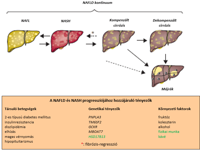
Factors written in black contribute to the progression of NAFLD and NASH; those written in green are protective factors.
NAFL: non-alcoholic fatty liver; NAFLD: non-alcoholic fatty liver disease; NASH: non-alcoholic steatohepatitis;
According to current epidemiological data, prevalence ranges from 13.8% (Africa) to 31.8% (Middle East). The difference can be explained by calorie intake, physical activity, body fat distribution, socioeconomic status and genetic characteristics. There is a close association between the disease and metabolic syndrome: the condition is seen in 47.3–63.7% of people with type 2 diabetes and in around 80% of people with obesity. Nevertheless, it can also occur with a healthy BMI (white people: <25 kg/m2, Asian people: <23 kg/m2) (non-obese NAFLD). Cirrhosis or liver cancer develops in fewer than 10% of patients, 10–20 years after diagnosis. Liver disease of other origin (alcohol-related, due to viral infection or autoimmune) has a synergistic effect on the condition. Mortality among NAFLD patients is only slightly higher than in the general population. In most cases the disease is stable or progresses slowly and does not lead to cirrhosis or liver-related death. Fibrosis can develop in both NAFL and NASH patients, but it progresses faster in the NASH group. The direct way of assessing fibrosis is histological evaluation of a liver biopsy sample, on the basis of which stages 0 (no fibrosis) to 4 (cirrhosis) can be determined 3,28. Determining the degree of fibrosis is the main prognostic parameter for estimating liver-related outcomes. At the cirrhosis stage the typical clinical picture of NASH is no longer visible, so when the condition cannot be explained by another disease (cryptogenic cirrhosis) and is associated with metabolic disease, it is referred to as "burnt-out" NASH. It is not uncommon for NASH to be recognised only once portal hypertension (with an enlarged spleen and a low platelet count) or another liver-related complication has developed. Around 3–4% of compensated cirrhoses become decompensated each year (ascites, hepatic encephalopathy, oesophageal and gastric bleeding)3. Liver cancer develops from NASH cirrhosis in 10.6/1,000 cases, while around 20% of NAFLD-related liver cancers develop from non-cirrhotic disease 3. The incidence of hepatocellular cancer of non-cirrhotic origin is low: 0.08/1,000 person-years. NAFLD is the leading cause of end-stage liver disease and the second most common cause of primary liver cancer3,30.
Apart from progressive liver disease, the leading cause of death in NAFLD is cardiovascular disease, followed by extrahepatic malignancies (bowel cancer or breast cancer). The link between cardiovascular disease and NAFLD is not fully understood, but the main factors most likely include metabolic syndrome, hypertension and an atherogenic lipid profile. Cancer (gastrointestinal, liver, uterine) is 1.9 times more common in NAFLD patients, probably owing to visceral adiposity and chronic low-grade inflammation.
Pathogenesis
The main pathogenetic factor in NAFLD is overnutrition. Unchanged fat uptake by liver cells due to insulin resistance and increased de novo lipogenesis overwhelm the metabolic capacity of the liver. The disruption of lipid metabolism leads to the formation of lipotoxic lipids, which contributes to cellular stress, inflammasome activation, apoptotic cell death, subsequent inflammation, tissue regeneration and fibrogenesis. These processes are made up of several metabolic, genetic and microbiome-dependent factors that are not yet precisely understood. Genetic factors account for 20–70% of the risk of the disease. The best-characterised SNP involved in the pathogenesis of NAFLD is a variant of the PNPLA3 gene. The SNPs known so far account for only 10–20% of the heritable predisposition. Metabolites derived from an altered gut microbiome contribute to fibrosis and inflammation (lipopolysaccharides, short-chain fatty acids, bile acid metabolites)3,35.
Estimating the severity of the disease
A diagnosis of NAFLD is most often suggested by imaging, by clinical risk scores (e.g. the fatty liver index) or during histological evaluation of a biopsy sample. Non-alcoholic fatty liver is most often first suspected during an abdominal ultrasound examination: a bright liver echotexture and blurred vessels are visible. Ultrasound has two limitations: 1) advanced fibrosis makes the echotexture coarser and the vessels blurred, and 2) the method is not sensitive enough – only steatosis above 30% can be detected. MRI-based measurement of steatosis (MRI proton density fat fraction) is much more sensitive (it detects steatosis above 5%), but it is currently used only for research purposes and for evaluating NASH treatment.
Type 2 diabetes (T2DM) is a major risk factor for progressive disease: it is associated with a twofold risk of fibrosis, cirrhosis complications and liver-related death3,16,54. Obesity (BMI > 30 kg/m2), lipid abnormalities (low HDL cholesterol, high triglycerides) and high blood pressure are also risk factors, but their effect is smaller than that of T2DM. Fibrosis is more pronounced in people over 60 than in younger people. The PNPLA3 gene variant is associated with a more severe histological picture, a higher probability of developing hepatocellular carcinoma, and higher liver-related and all-cause mortality3. Fasting insulin levels measured by mass spectrometry can predict NAFLD occurring without T2DM24. Modern diagnostics is also trying to bring metabolomic and lipidomic measurements into use25.
Several risk models exist for predicting T2DM. Prediabetes risk scores take the following parameters into account: sex, age, BMI, waist circumference, family history, ethnicity, systolic and diastolic blood pressure, heart rate, cholesterol, triglycerides, HDL, fasting blood glucose and HbA1c10b.
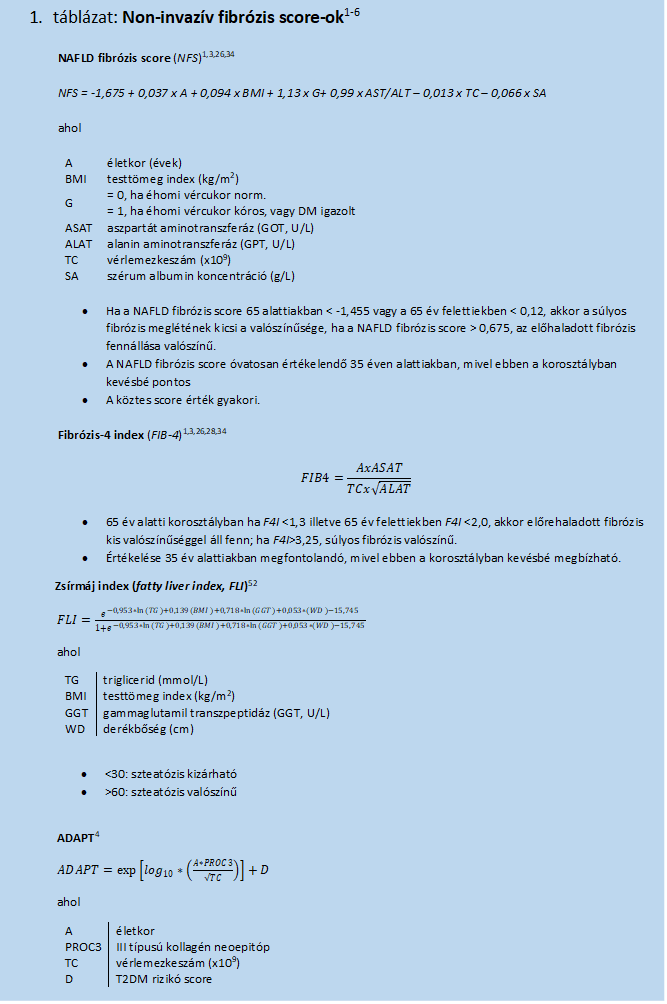
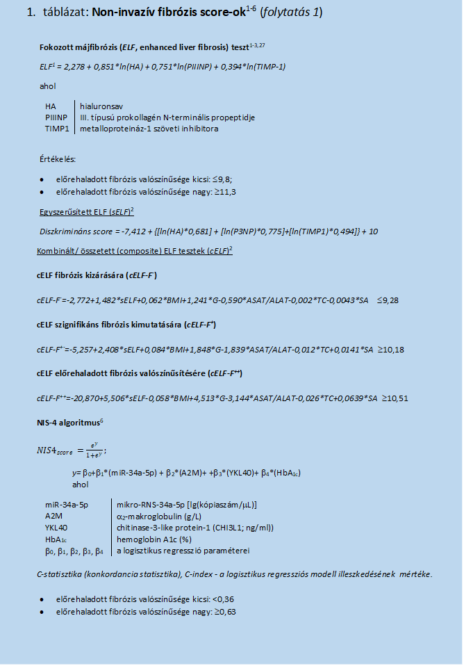
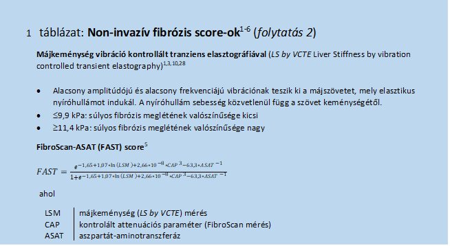
Clinicians often use indices derived from the results of non-invasive tests to assess the severity of the disease (Table 1). Classically, histological evaluation of a liver biopsy (determining the degree of steatosis, inflammation, hepatocyte ballooning and fibrosis) would be the reliable way of making the diagnosis, but it is an invasive method and is not free from intra- and inter-observer variability3,25. The degree of fibrosis correlates best with liver-related morbidity and mortality. Among the non-invasive methods, the NAFLD fibrosis score (NFS) and the fibrosis-4 index (FIB-4) have a sufficiently high negative predictive value (for ruling out advanced fibrosis)3. Further indices that can be calculated from laboratory markers – the Enhanced Liver Fibrosis (ELF)1,3 and composite ELF (cELF)2 scores, the ADAPT index4 supplemented with ProC3 measurement (a neoantigen generated by protease cleavage of type III collagen during the development of fibrosis4b), and the NIS4 algorithm6 (NIS4) – achieve better sensitivity and a larger area under the ROC (receiver operating characteristic) curve (AUROC).
Another group of methods for estimating fibrosis is the assessment of liver stiffness. The most widespread of these is the measurement of liver stiffness by vibration-controlled transient elastography (LS by VCTE); this parameter can also be estimated with two-dimensional shear wave elastography and MR elastography3,15,22,23,29. Together with liver stiffness, steatosis can also be assessed by measuring the controlled attenuation parameter (FibroScan). The values obtained correlate well not only with the risk of liver cirrhosis complications but also with the risk of developing liver cancer. Serum iron levels can also help in monitoring the risk of hepatocellular carcinoma33. Further extending these parameters either allows a more sensitive estimate (FAST score) or helps decide whether a therapeutic intervention is needed (Baveno VI criteria: LS by VCTE ≥20 kPa + platelet count <150*109 → varices requiring treatment)3,5. Another laboratory parameter for predicting liver fibrosis is growth differentiation factor-15 (GDF15)20. The adipokine isthmin improves glucose tolerance and hepatic steatosis21, and its level probably correlates with stable, non-progressive disease.
Prevention of the disease
Prevention is particularly important, given that few treatment options are currently available. The main means of prevention are improving diet quality and maintaining or increasing physical activity.
In people with obesity, type 2 diabetes or metabolic syndrome, it is essential to assess NAFLD/NASH/liver fibrosis using a non-invasive method. If a diagnosis of NASH is confirmed, besides ruling out advanced fibrosis and cirrhosis, cardiovascular risk factors and other comorbidities also need to be mapped.
If non-alcoholic fatty liver is suspected, it is advisable first to use simple non-invasive methods (NFL, FIB4) to rule out advanced fibrosis. Those in the negative group should be followed up (the measurements should be repeated at least every 3–5 years). If, on the basis of these parameters, the patient falls into the intermediate or probable advanced fibrosis group, further tests (elastography, ELF, NIS4, ADAPT, FAST) become necessary. Because the prevalence of the disease is particularly high among people with type 2 diabetes (40–70%), and because advanced fibrosis occurs in 10–20% of diabetes patients under care, they need closer follow-up and regular multidisciplinary monitoring of comorbidities 3,31,32,53,57,60
Treatment of non-alcoholic fatty liver
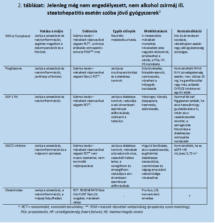
The cornerstones of treatment are the good-quality diet already mentioned under prevention50,55, plenty of fluids51 and physical activity – these are fundamental to improving metabolic syndrome and reducing body weight. In severe obesity, weight-loss surgery may also be considered44,45 . No medicine approved by the FDA (Food and Drug Administration, US) or the EMA (European Medicines Agency) is currently available. Studies of several investigational substances have not yet been completed; they are summarised in Table 23. The conditions for properly evaluating agents intended to treat NASH can be found in the articles by J V Lazarus and colleagues47,53. They also emphasise the importance of taking sex differences into account in the evaluation19. The endpoints of the studies were defined either as improvement of steatohepatitis without worsening of fibrosis, or as improvement of fibrosis without worsening of steatohepatitis3,47. The medicines studied include RRR-α-tocopherol, part of the vitamin E complex (see appendix); the thiazolidinedione pioglitazone, used in the treatment of T2DM; GLP-1 receptor agonists (semaglutide, liraglutide); SGLT2 inhibitors (canagliflozin, dapagliflozin, empagliflozin)17,56,58; obeticholic acid; and the combination of cilofexor and firsocostat3. Trials are under way with the pan-PPAR agonist lanifibranor36,37 and with Terns Pharmaceuticals' farnesoid X receptor agonist known as TERN-10138. In theory, agents acting on other targets (e.g. galectin-346) have also been proposed.
Other: in the US, a dietary supplement called LiverLuv is being tried (see appendix). Favourable effects of coffee consumption and of short-chain fatty acids (SCFAs) have also been observed48,49,59.
References
1. Q M Anstee, E J Lawitz: Noninvasive Tests Accurately Identify Advanced Fibrosis due to NASH: Baseline Data From the STELLAR Trials, Hepatology 2019, 70(5):1521-1530, doi: 10.1002/hep.30842
2. V Nobili, J Parkes et al.: Performance of ELF Serum Markers in Predicting Fibrosis Stage in Pediatric Non-Alcoholic Fatty Liver Disease 2009, 136:160-167, 10.1053/j.gastro.2008.09.013
3. E E Powell, V Wai-Sun Wong, M Rinella: Non-alcoholic fatty liver disease 2021, https://doi.org/10.1016/ S0140-6736(20)32511-3
4. S J Daniel, D J Leeming et al.: ADAPT: An algorithm incorporating PRO-C3 accurately identifies patients with NAFLD and advanced fibrosis 2019, 69(3):1075-1086, doi: 10.1002/hep.30163
4b M Boyle, D Tiniakos et al.: Performance of the PRO-C3 collagen neo-epitope biomarker in non-alcoholic fatty liver disease 2019, JHEP
Reports, https://doi.org/10.1016/j.jhepr.2019.06.004
5. P N Newsome, M Sasso et al: FibroScan-AST (FAST) score for the non-invasive identification of patients with non-alcoholicsteatohepatitis with significant activity and fibrosis: a prospective derivation and global validation study 2020, Lancet Gastroenterology and Hepatology, 5(4):362-373, doi: 10.1016/S2468-1253(19)30383-8
6. S A Harrison, V Ratziu et al.: A blood-based biomarker panel (NIS4) for non-invasive diagnosis of non-alcoholic steatohepatitis and liver fibrosis: a prospective derivation and global validation study 2020, https://doi.org/10.1016/S2468-1253(20)30252-1
7. 7 J Semmler, S Wernly et al: Metabolic Dysfunction-Associated Fatty Liver Disease (MAFLD)—Rather a Bystander Than a Driver of Mortality 2021, JCEM, 106(9):2670-2677, doi:10.1210/clinem/dgab339
8. M D Muthiah, N C Han, A J Sanyal: A clinical overview of non-alcoholic fatty liver disease: A guide to diagnosis, the clinical features, and complications—What the non-specialist needs to know 2021, Diabetes Obes Metab, DOI: 10.1111/dom.14521
9. J H Shubrook: Fatty Liver: Managing a Silent Epidemic in Primary Care 2021, Medscape, https://www.medscape.com/viewarticle/958321
10. W Pass: AGA Clinical Care Pathway: Screening, Diagnosis, and Treatment of NAFLD and NASH 2021, Medscape, https://www.medscape.com/viewarticle/963308
10b A Martinez-Millana, M Argente-Pla et al: Driving Type 2 Diabetes Risk Scores into Clinical Practice: Performance Analysis in Hospital
Settings 2019, J Clin Med, 8:107-126, doi:10.3390/jcm8010107
11. T Brand, I C Lamberta van den Munckhof et al: Superficial vs Deep Subcutaneous Adipose Tissue: Sex-Specific Associations With Hepatic Steatosis and Metabolic Traits 2021, JCEM 106(10):e3881-e3889, doi:10.1210/clinem/dgab426
12. G Targher, K E Corey et al: The complex link between NAFLD and type 2 diabetes mellitus — mechanisms and treatments 2021, Nature Rviews Gastroenterology & Hepatology, https://doi.org/10.1038/s41575-021-00448- y
13. D Barb, E M Repetto et al: Type 2 diabetes mellitus increases the risk of hepatic fibrosis in individuals with obesity and nonalcoholic fatty liver disease 2021, Obesity, 29:1950-1960, DOI: 10.1002/oby.23263
14. H Yki-Järvinen, P K Luukkonen et al: Dietary carbohydrates and fats in nonalcoholic fatty liver disease 2021, Nature Review Gastroenterology & Hepatology, https://doi.org/10.1038/s41575-021-00472- y
15. A Andersson, M Kelly et al: Clinical utility of MRI biomarkers for identifying NASH patients at high risk of progression: A multi-center pooled data and meta-analysis 2021, Clinical Gastroenterology and Hepatology, https://doi.org/10.1016/j.cgh.2021.09.041
16. C C Cohen, W Perng et al: Hepatic Fat in Early Childhood Is Independently Associated With Estimated Insulin Resistance: The Healthy Start Study 2021, JCEM 106(11):3140-3150, https://doi.org/10.1210/clinem/dgab541
17. P Borodavkin, W Sheridan et al: Effects of glucagon-like peptide-1 receptor agonists on histopathological and secondary biomarkers of non-alcoholic steatohepatitis: A systematic review and meta-analysis 2021, Diabetes Obes Metab 2021, DOI: 10.1111/dom.14565
18. S Kawata, A Nakamura et al: Glucokinase activation leads to an unsustained hypoglycaemic effect with hepatic triglyceride accumulation in db/db mice 2021, Diabetes Obes Metab, DOI: 10.1111/dom.14586
19. P Lefebre, B Staels: Hepatic sexual dimorphism — implications for non- alcoholic fatty liver disease 2021, https://doi.org/10.1038/s41574-021-00538-6
20. J Bilson, E Scorletti et al: Growth differentiation factor-15 and the association between type 2 diabetes and liver fibrosis in NAFLD 2021, Nutrition & Diabetes 11:32, https://doi.org/10.1038/s41387-021-00170-3
21. Z Jiang, M Zhao et al: Isthmin-1 is an adipokine that promotes glucose uptake and improves glucose tolerance and hepatic steatosis 2021, 33:1836-1852, https://doi.org/10.1016/j.cmet.2021.07.010
22. A Andersson, M Kelly et al: Clinical Utility of MRI Biomarkers for Identifying NASH Patients at High Risk of Progression: A Multicenter Pooled Data and Meta-Analysis 2021, Clinical Gastroenterology and Hepatology, https://doi.org/10.1016/j.cgh.2021.09.041
23. D Swift: Andrea Dennis on MRI Biomarkers for Identifying Progression to NASH – Quantitative MRI-based tests are non-invasive and effective 2021, MesPage Today, https://www.medpagetoday.com/reading-room/aga/lower-gi/95233
24. F Bril, M J McPhaul et al: Intact Fasting Insulin Identifies Nonalcoholic Fatty Liver Disease in Patients Without Diabetes 2021, JCEM 106(11):e4360-e4371, doi:10.1210/clinem/dgab417
25. M Masoodi, A Gastaldelli et al: Metabolomics and lipidomics in NAFLD: biomarkers and non-invasivediagnostic tests 2021, Nature Review Gastroenterology and Hepatology 18:835-856, https://doi.org/10.1038/s41575-021-00502-9
26. A Drolz, S Wolter et al: Performance of non-invasive fibrosis scores in non-alcoholic fatty liver disease with and without morbid obesity 2021, International Journal of Obesity, 45:2197-2204, https://doi.org/10.1038/s41366-021-00881-8
27. Z M Younossi, S Felix et al: Performance of the Enhanced Liver Fibrosis Test to Estimate Advanced Fibrosis Among Patients With Nonalcoholic Fatty Liver Disease 2021, JAMA Network Open, 4(9):e2123923. doi:10.1001/jamanetworkopen.2021.23923
28. T Poynard, V Paradis et al: Prospective External Validation of a New Non-invasive Test for theDiagnosis of Non-alcoholic Steatohepatitis in Patients With Type 2Diabetes 2021, Aliment Pharmacol Ther., 54(7):952-966, https://www.medscape.com/viewarticle/958927
29. N Tamaki, V Ajmera, R Loomba: Non-invasive methods for imaging hepatic steatosis and their clinicalimportance in NAFLD 2022, Nature Reviews Endocrinology, 18:55-66, https://doi.org/10.1038/s41574-021-00584-0
30. A J Sanyal, M L Van Natta et al: Prospective Study of Outcomes in Adults with Nonalcoholic Fatty Liver Disease 2021, N Engl J Med, 385:1559-1569, DOI: 10.1056/NEJMoa2029349
31. G Zuo, L Xuan et al: New Nonalcoholic Fatty Liver Disease and Fibrosis Progression Associate With the Risk of Incident Chronic Kidney Disease 2021, JCEM, 106(10):e3957-e3968, https://doi.org/10.1210/clinem/dgab425
32. S A Polyzos, S Kechagias, E A Tsochatzis: Non-alcoholic Fatty Liver Disease and Cardiovascular Diseases 2021, Aliment Pharmacol Ther., 54(8):1013-1025, https://www.medscape.com/viewarticle/960741
33. Z Hamza, S Writer: Clinical surveillance of serum iron levels may be worth monitoring, researcher says, 2021 MedPage Today https://www.medpagetoday.com/meetingcoverage/aasld/95645
34. T J Oh, K Kim et al: Liver Fibrosis Indices for the Prediction of Mortality in Korean Subjects: A 16-Year Prospective Cohort Study 2021, 10:1-8, https://doi.org/10.1210/jendso/bvab123
35. J I Martínez-Montoro, M S Kuchay et al: Gut microbiota and related metabolites in the pathogenesis of nonalcoholic steatohepatitis and its resolution afterbariatric surgery 2021, Obesity Reviews, https://doi.org/10.1111/obr.13367
36. S M Francque, P Bedossa et al: A Randomized, Controlled Trial of the Pan-PPAR Agonist Lanifibranor in NASH 2021, N Engl J Med, 385:1547-1558, DOI: 10.1056/NEJMoa2036205
37. M Brooks: Novel Drug Lanifibranor Promising for Nonalcoholic Steatohepatitis 2021, ttps://www.medscape.com/viewarticle/961293
38. R Loomba, A K Singal: Data on TERN-101 Show Promise for NASH Treatment 2021, Gastroenterology & Endoscopy News 2021, https://www.gastroendonews.com/Article/PrintArticle?articleID=64811
39. A Aminian, A Al-Kurd et al: Association of Bariatric Surgery With Major Adverse Liver and Cardiovascular Outcomes in Patients With Biopsy-Proven Nonalcoholic Steatohepatitis 2021, JAMA, 326(20):2031-2042. doi:10.1001/jama.2021.19569
40. K E Corey, Z N Memel: Bariatric Surgery as a Strategy for Improving Outcomes in Nonalcoholic Steatohepatitis 2021, JAMA 326(20):2015-2017. doi:10.1001/jama.2021.17451
41. K A Seeberg, H Borgeraas et al: Do Gastric Bypass and Sleeve Gastrectomy Help Reduce Liver Fat Content and Scarring in Patients With Type 2 Diabetes? 2022, https://doi.org/10.7326/P21-0012
42. N Finer: Weight loss interventions and nonalcoholic fatty liver disease: Optimizing liver outcomes 2021, Diabetes Obes Metab, DOI: 10.1111/dom.14569
43. P Harrison: Weight Loss Surgery ‘Best Treatment’ for Fatty Liver Disease 2021, Medscape, https://www.medscape.com/viewarticle/963529
44. F Salomone, W Currenti et al: Effects of Intragastric Balloon in Patients With Non-alcoholic FattyLiver Disease and Advanced Fibrosis 2021, Liver International 41(9):2112-2116, https://www.medscape.com/viewarticle/957258
45. K A Seeberg, H Borgeraas et al: Gastric Bypass Versus Sleeve Gastrectomy in Type 2 Diabetes: Effects on Hepatic Steatosis and Fibrosis, 2022, Annals of Internal Medicine, https://doi.org/10.7326/M21-1962
46. R E Soccio: Galectin-3 in NAFLD: Therapeutic Target or Noncausal Biomarker? 2021, JCEM, 106(9):e3773-e3774, doi:10.1210/clinem/dgab363
47. J V Lazarus, Q M Anstee et al: Defining comprehensive models of care for NAFLD 2021, Nature Review Gastroenterology & Hepatology, https://doi.org/10.1038/s41575-021-00477-7
48. S Niezen, M Mehta et al: Coffee Consumption Is Associated With Lower Liver Stiffness: A Nationally RepresentativeStudy 2021, Clinical Gastroenterology and Hepatology, https://doi.org/10.1016/j.cgh.2021.09.042
49. Z Hamza: A Cup of Joe to Thwart Liver Fibrosis? How About Four — Study finds protective benefit with high consumption, though not for fatty liverdisease 2021, MedPage Today, https://www.medpagetoday.com/gastroenterology/generalhepatology/94940?xid=nl_mpt_DHE_2021-10-09&eun=g1487684d0r&utm_source=Sailt%E2%80%A6
50. L Valenti: Food Concerns Linked to Liver Disease 2021, Gastroenterology & Endoscopy News 2021, https://www.gastroendonews.com/Article/PrintArticle?articleID=64810
51. X Wang, S Lin et al: Higher plain water intake is related to lower newly diagnosed nonalcoholic fatty liver disease risk: a population-based study 2021, European Journal of Clinical Nutrition, https://doi.org/10.1038/s41430-021-00891-9
52. A nem alkoholos zsírmáj kockázatának felbecslése 2022, Medical Online,
http://medicalonline.hu/gyogyitas/cikk/a_nem_alkoholos_zsirmaj_kockazatanak_felbecslese
53. J V Lazarus, H E Mark et al: Advancing the global public health agenda for NAFLD: a consensus statement 2022,Nature Reviews Gastroenterology & Hepatology, 19:60-78, https://doi.org/10.1038/s41575-021-00523-4
54. X Hu, W Yu et al.: First-degree family history of diabetes is associated with nonalcoholic fatty liver diseaseindependent of glucose metabolic status 2021, Journal of Diabetes and Its Complications, 36(1), https://doi.org/10.1016/j.jdiacomp.2021.108083
55. L Bosembiante, G Targher, C Maffeis: Non-alcoholic fatty liver disease in obese children and adolescents: a role for nutrition? 2022, European Journal of Clinical Nutrition, 46:28-39, https://doi.org/10.1038/s41430-021-00928-z
56. A Mantovani, C D Byrne, G Targher: Efficacy of peroxisome proliferator-activated receptor agonists, glucagon-like peptide-1 receptor agonists, or sodium-glucose cotransporter-2 inhibitors for treatment of non-alcoholic fatty liver disease: a systematic review 2022, Lancet Gastroenterology & Hepatology, https://doi.org/10.1016/S2468-1253(21)00261-2
57. M M Ruissen, A L Mak et al: Non-alcoholic fatty liver disease: a multidisciplinary approach towards a cardiometabolic liver disease 2020, European Journal of Endocrinology, 183:R57-R73, https://eje.bioscientifica.com/view/journals/eje/183/3/EJE-20-0065.xml
58. R M Pontes-da-Silva, T de Souza Marinho et al: Obese mice weight loss role on nonalcoholic fatty liver disease and endoplasmic reticulum stress treated by a GLP-1 receptor agonist 2021, International Journal of Obesity, https://doi.org/10.1038/s41366-021-00955-7
59. M Deng, F Qu et al: SCFAs alleviated steatosis and inflammation in mice with NASH induced by MCD 2020, Journal of Endocrinology, 245:425-437, https://doi.org/10.1530/JOE-20-0018
60. G E Chung, E J Chou et al: Young adults with nonalcoholic fatty liver disease, defined using the fatty liver index, can be at increased risk of myocardial infarction or stroke 2021, Diabetes Obes Metab, DOI: 10.1111/dom.14597
61. N Chalasani, Z Younossi et al: The diagnosis and management of nonalcoholic fatty liver disease: Practice guidance from the American Association for the Study of Liver Diseases 2018, Hepatology, 67(1):328-357, doi: 10.1002/hep.29367
Appendix: vitamin E, LiverLuv
Vitamin E
Vitamin E is in fact a mixture of several compounds: the tocopherols (α, β, γ, δ) and the tocotrienols (α, β, γ, δ). The name tocopherol is made up of the Ancient Greek words τοκοσ (birth) and Φερειν (to carry) ("protector of pregnancy"), while the ending -ol refers to the
–OH group in the molecule.
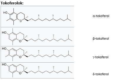
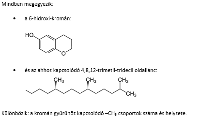
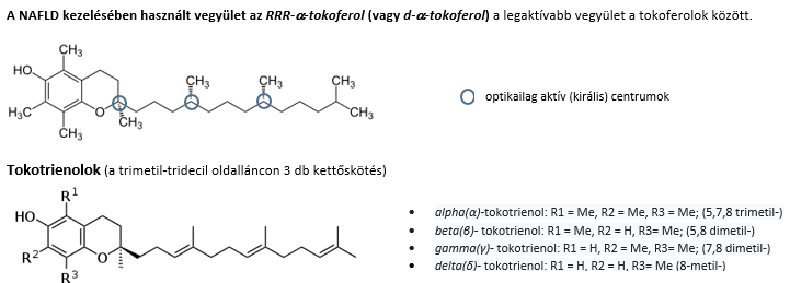
Vitamin E (a mixture of tocopherols/tocotrienols) is a fat-soluble vitamin and an effective free radical scavenger (antioxidant). The recommended dietary allowance (RDA) is 15 mg (calculated as α-tocopherol). This is equivalent to 22.5 IU (since 1 mg=1.5 IU). Because it is a fat-soluble vitamin, the maximum daily intake is limited. The European Food Safety Authority (EFSA) has set the maximum daily intake, calculated as α-tocopherol, at 300 mg (450 IU). The main dietary sources of vitamin E are vegetable oils pressed from oilseeds.
Vitamin E deficiency is rare; when it does occur, problems with nerve conduction and neuromuscular transmission, spinocerebellar ataxia and myopathies may develop. Because the vitamin is an antioxidant, its deficiency may also cause anaemia due to oxidative damage to red blood cells. Its deficiency makes Alzheimer's disease or vascular dementia more likely. Its effect in preventing tumour formation, cataracts and cardiovascular disease has not been clearly proven. Likewise, there is no clear evidence that it protects against obstetric risk factors (preterm birth, pre-eclampsia, neonatal death).
High doses of vitamin E potentiate the effect of aspirin and warfarin and lower the serum levels of tamoxifen and ciclosporin A. High doses increase the risk of bleeding and of cardiovascular events61.
LiverLuv
Website: https://luvbynature.com/liver-luv/?AFFID=402363
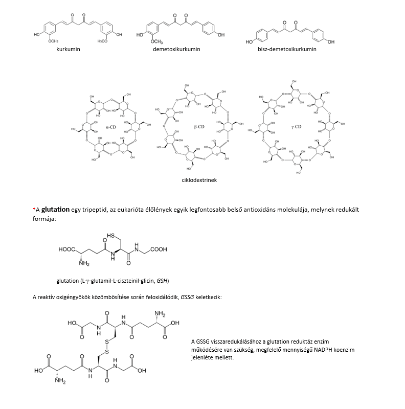
Several American hepatologists recommend this liver-protecting, antioxidant preparation (dietary supplement), made mainly from herbal extracts (glutathione* supplements). The active ingredients are packaged in liposomes containing phosphatidylcholine, which ensures that they pass through the cell membrane and allows the molecules delivered in this way to act inside the cell.
Composition:
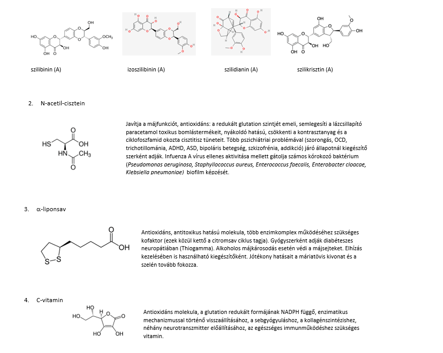
1. Milk thistle extract (liver-protecting, detoxifying, antioxidant)
The plant's main active compound is silymarin, a mixture of several flavonolignans, whose most important components are:
5. Selenium
A trace element with anti-tumour and heart-muscle-protecting effects. It is a component of several important enzymes, e.g. glutathione peroxidase, which has an antioxidant role and is important for the integrity of subcellular membranes; thioredoxin reductase, which is needed to restore the reduced form of glutathione; and the deiodinases required for thyroid hormone metabolism.
6. Turmeric extract
The plant's main active compounds, the curcuminoids, are also antioxidants – compounds that can effectively neutralise superoxide radicals – and are present in the plant as complexes with cyclodextrin.
SIBO and food intolerances: is food necessarily to blame?
Bloating after milk, bread or onions? The way the digestive system works may also play a role behind these symptoms. Dr Anikó Hovanecz’s article expla...

A corporate health programme in practice – the SFC CEO’s experience
What does caring for employees and their family members mean for a company leader? Through his personal experience, Gábor Horváth, CEO of SFC, explain...

Don't just get the school bag ready
Starting school is not only about new exercise books and a changed daily routine but also about reorganising family meals. The FOODTEST Children's pac...