Author: Dr Károly Krkos
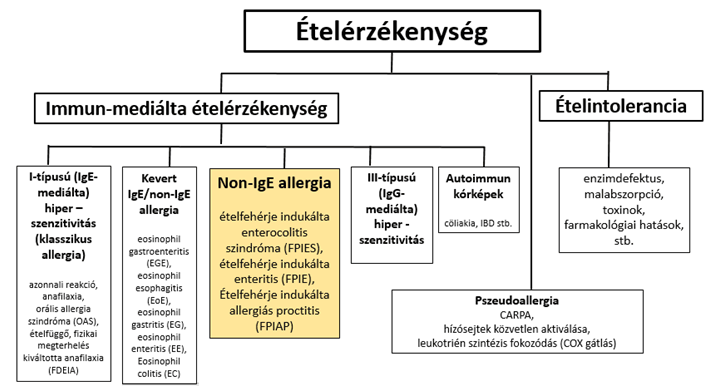
Food sensitivities are usually divided into two large groups: conditions that cause gastrointestinal and other symptoms without the involvement of the immune system (intolerances), and disease processes involving the immune system (immune-mediated food sensitivities). The former are caused by malabsorption, enzyme defects, conditions due to toxic contamination and pharmacological effects2. In a large proportion of immune-mediated food sensitivities, IgE antibodies play the decisive role (classic allergy, type I hypersensitivity). There are, however, conditions in which the decisive role is played not by IgE production and mast cell activation, but by other mechanisms that contribute to the pathogenesis with or without IgE formation ("mixed IgE/non-IgE", "non-IgE allergy"). So-called non-IgE allergies are characterised neither by IgE nor by IgG production. They differ from processes involving type III hypersensitivity (IgG-mediated food sensitivities). Pseudoallergies form a separate category; they are based on activation of the mast cell by a route other than IgE–FcεRI (complement activation-related pseudoallergy – CARPA, direct activation via other receptors, a predominance of leukotriene synthesis due to COX inhibition).
Figure 1: Classification of food sensitivities
In diagnosing mixed IgE/non-IgE and non-IgE allergies, the primary role is played by an accurate, detailed medical history and a thorough understanding of the course of the disease, which in many cases needs to be supplemented by imaging, endoscopic methods and examination of biopsy samples.
Non-IgE-mediated gastrointestinal food allergy (non-IgE-GI-FA) comprises three main conditions: food protein-induced enterocolitis syndrome (FPIES), food protein-induced enteropathy (FPE) and food protein-induced allergic proctocolitis (FPIAP)1,13,15. Two very rare conditions also belong here: Heiner syndrome (pulmonary haemosiderosis)13,20-23 and cow's milk-induced iron deficiency anaemia (cow's milk protein allergy-related iron deficiency anemia, CMPA-IDA)13,24,25.
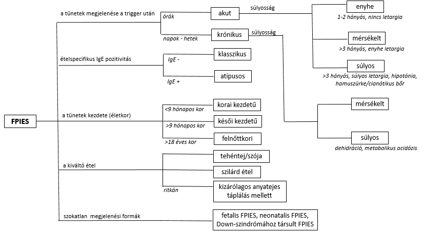
Whereas in classic, IgE-mediated food allergy (IgE-FA) the symptoms appear shortly after eating (1–2 minutes to 1 hour) and are not limited to the gastrointestinal tract (the skin, mucous membranes and possibly breathing and circulation are also affected, and anaphylaxis may develop), in the non-IgE-GI-FA conditions the symptoms appear later (after 2–3 hours) and are limited exclusively to the digestive system1,13,15. Typically, no food-specific IgE antibody can be detected (apart from a small proportion of FPIES cases)1,13-19. The exact pathomechanism is not known. Non-IgE-GI-FA diseases form a continuum. The expression and severity of the symptoms depend on which section(s) of the gastrointestinal tract are affected: in FPIAP, where the process is limited to the distal colon, fresh blood is passed from the rectum (haematochezia) in otherwise healthy-looking infants. In FPE, which involves the small intestine, malabsorption and failure to thrive (FTT) dominate the clinical picture. FPIES, the most severe form, often producing acute and alarming symptoms, indicates involvement of the entire gastrointestinal tract. Its main feature is intractable vomiting and, in more severe cases, metabolic disturbances and hypovolaemic shock. FPIES can be further classified by the timing of symptom onset (acute and chronic), severity (mild, moderate, severe), the patient's age (early onset, late onset, adult) and the type of trigger food (caused by cow's milk/soya or by solid food). Food-specific IgE antibodies can also be detected in some patients ("atypical FPIES" – Figure 2).
Figure: Classification of FPIES (based on L Labrosse, F Graham and J-C Caubet: Non-IgE-Mediated Gastrointestinal Food Allergies in Children: An Update)
The prevalence of non-IgE-GI-FA diseases has been little studied; FPIAP is considered the most common condition. 60% of infants examined for rectal bleeding turn out to have FPIAP. The prevalence of FPE is the least known, but it is certain that its incidence is declining, presumably because breastfeeding has become more popular and infant formulas have improved in quality. FPIES is a rare disease; based on surveys, its incidence is estimated at 0.3–0.7%. This group of diseases is often associated with an atopic condition (40–60% of patients with FPIES are affected, and 40–50% of those with FPE and FPIAP), and 60–80% of family members are atopic.
Atopy26-28 is a genetically linked susceptibility to allergic diseases: allergic asthma, dermatitis, rhinitis (hay fever) and food allergy. This group of diseases is characterised by the so-called atopic march: initial atopic dermatitis (and food allergy) is followed by allergic asthma and rhinitis. The dermatitis–asthma–rhinitis trio is also called the atopic triad.
Heiner syndrome20-23 is an atypical lung disease induced by cow's milk protein in infants and young children. It is characterised by recurrent respiratory symptoms, infiltrates on chest X-ray mimicking pneumonia, fever, anaemia and failure to thrive. In some children, gastrointestinal symptoms (vomiting, diarrhoea, abdominal pain, bloody stools) also appear in addition to those listed above. Its pathology is not clear, but type III hypersensitivity (immune complex formation) is thought to be behind it. The symptoms appear 1–9 months after starting a diet containing cow's milk. In about 10% of cases the disease is severe and leads to pulmonary haemosiderosis. Enlargement of the liver, spleen and lymph nodes is occasionally observed. There have been cases where, alongside marked gastrointestinal symptoms, haematochezia (passing blood through the rectum) dominated the clinical picture21. Haemosiderin-laden macrophages are seen in bronchial and gastric aspirates. In many cases, IgG positivity against milk protein can be detected. IgE is negative in most cases, but positivity may also occur. The symptoms resolve 5–21 days after dairy products are withdrawn. In its severe form, the disease can lead to alveolar hypoventilation, massive acute pulmonary haemosiderosis, pulmonary hypertension and cor pulmonale, which is why early recognition of the condition and elimination of cow's milk from the diet are important.
The most common cause of anaemia in infancy and early childhood is iron deficiency (its prevalence between 1 and 3 years of age is estimated at 0.9–4.4%). Its main cause is inadequate iron intake. In a small percentage of cases, however, the iron deficiency is caused by increased losses due to cow's milk sensitivity: this is iron deficiency anaemia associated with cow's milk allergy (cow's milk allergy-related iron deficiency anemia, CMA-IDA)24,25. Its symptoms: pallor (100%), hypoalbuminaemia (86%), occult blood in the stool (57%), generalised oedema (43%), failure to thrive (43%). Four fifths of patients show positivity for IgE against cow's milk. On colonoscopy, erosive haemorrhagic colitis and lymphoid hyperplasia are seen, but no eosinophilia. The condition resolves with withdrawal of milk and iron supplementation.
The pathology of non-IgE-GI-FA is little explored1,3,16. An inadequate cellular immune response is considered the main cause of the disease. In FPIES, publications describe increased TNF secretion and reduced TGF secretion by food antigen-specific T cells. Local T-cell infiltration and inflammation lead to increased intestinal permeability and easier entry of antigens. A predominant Th2 response with IL-4, IL-5, IL-9 and IL-13 production is linked to atopic predisposition. During an oral food challenge (OFC), cells of the innate immune system (monocytes, neutrophils, eosinophils and NK cells) are activated. IL-9 production results in mast cell recruitment and intestinal anaphylaxis. Baseline tryptase levels are higher in OFC-positive individuals – showing that mast cells also play a significant role in the process. Studies of humoral immunity found low levels of neutralising specific IgG4 antibodies.
In FPE, damage to the structure of the jejunal mucosa has been observed, with food-specific (mainly cow's milk-specific) T-cell infiltration, leading to malabsorption. A leading role in the development of the disease is attributed to the activation of cytotoxic (CD8+) T cells. Large numbers of γδ-TCR+ cells between the absorptive epithelial cells have also been observed.
The main pathological feature of FPIAP is eosinophilic infiltration of the rectosigmoid mucosa. This condition develops mainly in breastfed infants, so it is assumed that components in breast milk that trigger an immune reaction may play a role in its development.
Based on clinical factors, Table 1 below shows the characteristics of the 3 symptom groups (and the FPIES subgroups) (based on 1. L Labrosse, F Graham and J-C Caubet: Non-IgE-Mediated Gastrointestinal Food Allergies in Children: An Update; abbreviations: A=acute, C=chronic, OFC=oral food challenge):
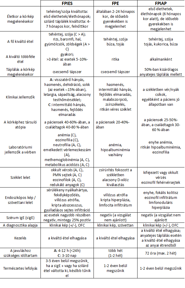
A= acute; C= chronic; PMN cells= polymorphonuclear cells
The leading symptom of acute FPIES1,13-15,17 is repeated vomiting appearing 1–4 hours after eating (in more than 95% of cases). Diarrhoea appears 5–10 hours after eating, in a smaller proportion (25–50% of patients). In newborns, symptoms resembling sepsis can also be observed: lethargy (65–100%), pallor (30–90%), hypothermia (5%)7. The clinical picture can also be severe: haemodynamic instability develops in 15%.
Chronic FPIES1,13-15,17 is characterised by persistent (sometimes mucous and bloody) diarrhoea, accompanied by intermittent vomiting, abdominal distension and failure to gain weight. In a group of patients, gradual worsening of the symptoms leads to dehydration (15–45%) and metabolic disturbances (5%)29-31. Whereas in chronic FPIES the trigger food is cow's milk or soya, the acute form is more often provoked by the introduction of solid food (most often rice). FPIES occurs only in a small percentage of cases (0–5%) with exclusive breastfeeding. In recent years, adult FPIES has also been described, most often triggered by seafood3. A form of the disease appearing in the neonatal period has also been identified, caused by sensitisation in the womb4.
The symptoms of FPE1,13,15 resemble those of coeliac disease and in most cases appear in infancy, when cow's milk is introduced: chronic diarrhoea, malabsorption, fatty stools and failure to thrive (FTT), which improve when cow's milk is withdrawn. Vomiting may also occur. The extra-intestinal problems seen in coeliac disease (e.g. dermatitis herpetiformis) are not observed. Because of the mucosal damage, secondary carbohydrate intolerance also develops5.
Unlike the conditions mentioned above, FPIAP often occurs with exclusive breastfeeding. This is explained by antigens passing from the mother's diet into the breast milk32. Its main symptom is loose, bloody and possibly mucous stools. Otherwise the infant is healthy; there is no forceful vomiting, diarrhoea or failure to thrive. Food refusal and irritability can be observed in some patients. Although the disease is typically encountered in infancy, it can also occur in older children (in 18%)33.
Among the foods triggering non-IgE-GI-FA, cow's milk ranks first (in 40–50% of cases, and in 65% of children with FPIES13), but many other foods can also trigger it – these vary by condition and by country (and with eating habits and differing genetic background). Besides cow's milk, the main provoking food of FPIES is soya. In many cases the symptoms are triggered not by eating a single component, but by 2 (in 25% of cases13), 3 or even more (mainly with solid triggers – in fewer than one in ten cases13). Among solid foods, cereals (rice, oats), among vegetables sweet potato and squash, among fruits banana and avocado, as well as poultry and egg feature as triggers. In the United States and Australia, rice is the most common culprit and causes more severe symptoms7. In Italy and Spain, fish and seafood take first place. Among the triggers of FPE, cow's milk also leads, possibly together with soya, followed by wheat and, to a lesser extent, banana, egg and meat. Finally, FPIAP is characterised by sensitivity to cow's milk (and other foods – via breast milk, less often directly). (Other foods: soya, egg, wheat and maize.)
In diagnosing non-IgE-GI-FA, the leading role is played by careful analysis of the clinical symptoms and a food challenge carried out under hospital supervision (oral food challenge – OFC), except for FPE, which usually also requires histological examination for confirmation1,1,13-15-17,18.
The basics of diagnosis are summarised in Table 2 (based on 1. L Labrosse, F Graham and J-C Caubet: Non-IgE-Mediated Gastrointestinal Food Allergies in Children: An Update):
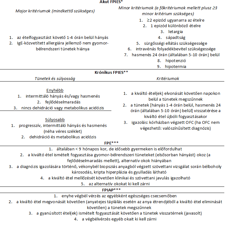
*: Both major criteria and at least 3 minor criteria must be met for the diagnosis **: General criteria owing to limited data (based on international consensus) ***: No defined set of criteria; features used in clinical practice (FPIAP: based on the EAACI position paper34)
The OFC (oral food challenge) is the gold standard for diagnosing FPIES and helps in differential diagnosis. The procedure carries a high risk, so it can only be carried out under hospital supervision (intravenous fluid replacement and correction of metabolic disturbance become necessary in about half of cases). During the challenge, which lasts half an hour, the patient receives the trigger food in 3 equal doses. This is followed by at least 4–6 hours of observation. For safety reasons, specific IgE testing before the challenge is advisable to rule out the possibility of classic allergy.
In the FPIES group, serum-specific IgE positivity occurs in a quarter of patients (atypical FPIES) – IgE measurement is not recommended in the other forms of the disease. The laboratory findings are not specific: anaemia, hypoalbuminaemia, raised platelet count (often with low mean platelet volume – MPV7), higher white blood cell count with a larger proportion of immature forms, and in more severe cases metabolic acidosis and methaemoglobinaemia may also develop. Typically, CRP is not raised or only minimally raised. A marked rise in CRP (>200mg/l) tends to rule out the condition, and an alternative diagnosis should then be considered7. FPE is characterised by the development of iron deficiency anaemia and hypoproteinaemia, whereas FPIAP is rarely associated with serum abnormalities. Neutrophil and eosinophil leucocytes, Charcot–Leyden crystals and reducing substances may appear in the stool35. In FPIES and FPIAP, occult bleeding and increased calprotectin excretion can occasionally be detected.
Imaging procedures are not among the basic diagnostic procedures in this group of diseases. X-ray examination sometimes shows gas formation within the bowel wall, which may mimic necrotising enterocolitis. Ultrasound may show thickening of the small bowel wall and slowed peristalsis, which disappear after the trigger food is withdrawn.
Endoscopic examinations are needed to diagnose FPE, in order to take biopsy samples. Jejunal villous atrophy and crypt hyperplasia milder than those seen in coeliac disease can be observed. The villus/crypt ratio is a sensitive indicator of damage to the jejunal mucosa6. On examination in FPIAP, hyperaemia of the mucosa with tiny haemorrhages is striking. The inflammation is accompanied by eosinophilic infiltration, lymphonodular hyperplasia and an increase in mast cells35.
The data needed to rule out other conditions (for differential diagnosis) are summarised in Table 3 below:
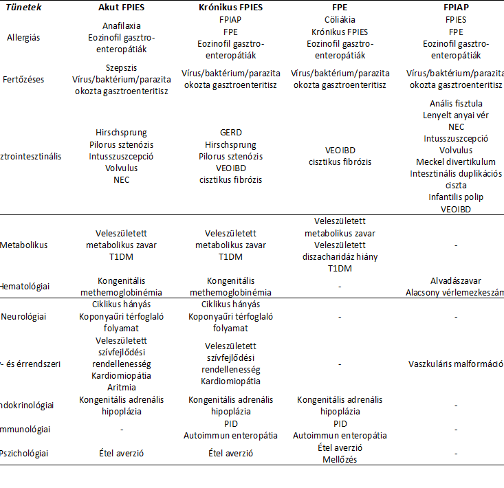
GERD: gastro-oesophageal reflux disease; NEC: necrotising enterocolitis; PID: primary immunodeficiency; T1DM: type 1 diabetes; VEOIBD: very early onset inflammatory bowel disease
The natural course of non-IgE-GI-FA is favourable in the majority of cases. Most children with onset in infancy outgrow the disease by school age. FPIES usually resolves before the age of 3–5 years, but the course of later-onset and atypical FPIES is more protracted. Moreover, the latter condition may turn into an immediate, IgE-mediated reaction (classic allergy)8, and the opposite can also occur: IgE-mediated food allergy may be replaced by non-IgE-GI-FA9,10. FPE typically resolves within 1–2 years (unlike coeliac disease, which it resembles), but rarely it may persist into late childhood11. FPIAP often resolves by 11 months of age and rarely persists until 1–2 years of age. Patients who have had FPIAP are more likely to develop functional gastrointestinal disorders later, especially those in whom FPIAP was more severe and more protracted12.
In treatment1,13,18, elimination of the trigger food plays a key role. The aim is not to eliminate foods unnecessarily, and if any dietary change is made, it should always be done under the supervision of a dietitian so that deficiency diseases can be avoided (particular attention must be paid to adequate vitamin D, calcium, zinc and selenium intake). Two approaches to food elimination are used: the "bottom-up" or the "top-down" method. In milder cases the former is used: first, elimination of the most likely food (e.g. cow's milk) is recommended. If this is not enough, further foods are tried one after another. In severe cases an elemental diet (containing amino acids) is set up, which excludes allergens, and after improvement different foods are gradually reintroduced one after another.
When a cow's milk-free diet is indicated, either an extensively hydrolysed formula (EHF) or an amino acid-based formula (AAF) is recommended. Patients with FPIES may become seriously ill and then require emergency care to correct disturbances of fluid and electrolyte balance and metabolic balance. Epinephrine (adrenaline) may only be needed in IgE-positive FPIES (atypical FPIES). Since a large proportion of FPIAP cases occur with breastfeeding, cow's milk and soya products need to be excluded from the mother's diet.
Since in the vast majority of cases the disease resolves within a few years, possible recovery is checked every 12–18 months with an OFC test carried out in hospital. To prevent FPIES, the introduction of less allergenic foods during weaning is recommended: among vegetables, broccoli, cauliflower, parsnip and squash, and among fruits, blueberries are advised. The gradual, step-by-step introduction of foods of different colours, flavours and textures is considered important.
References
- L Labrosse, F Graham and J-C Caubet: Non-IgE-Mediated Gastrointestinal Food Allergies in Children: An Update 2020, Nutrients, 12:2086, doi:10.3390/nu12072086
- S Waserman, P Bégin and W Watson: IgE-mediated food allergy 2018, Allergy, Asthma & Clinical Immunology 14(Suppl. 2):55, https://doi.org/10.1186/s13223-018-0284-3
- S Ichimura, H Kakita et al: A Rare Case of Fetal Onset, Food Protein-Induced Enterocolitis Syndrome 2019, Neonatology, DOI: 10.1159/000502717
- M R Faber, P Rieu et al: Allergic colitis presenting within the first hours of premature life 2005, Clinical Observations, DOI: 10.1080/08035250510039892
- N Iyngkaran, Z Abdin et al: Acquired carbohydrate intolerance and cow milk protein-sensitive enteropathy in young infants 1979, The Journal of Pediatrics, 95(3):373-378
- R McCalla, E Savilahti et al: Morphology of the Jejunum in Children with Eczema due to Food Allergy 1980, Allergy, 35:563-571
- E Lee, R H Barnes et al: Differentiating Acute Food Protein–Induced Enterocolitis Syndrome From Its Mimics: A Comparison of Clinical Features and Routine Laboratory Biomarkers 2019 J Allergy Clin Immunol Pract, https://doi.org/10.1016/j.jaip.2018.10.020
- A Kessel, I Dalal: The pendulum between food protein–induced enterocolitis syndrome and IgE-mediated milk allergy 2011, Acta Paediatrica, 100:e183-e185, DOI:10.1111/j.1651-2227.2011.02257.x
- C Banzato, G L Piacentini et al: Unusual shift from IgE-mediated milk allergy to food protein-induced enterocolitis syndrome 2013, Eur Ann Allergy Clin Immunol, 45(6):209-211
- H Duffy, M Egan: Development of food protein–induced enterocolitis syndrome (FPIES) to egg after immunoglobulin E–mediated egg allergy 2018, Annals of Allergy, Asthma, Immunology, doi: 1016/j.anai.2018.05.028
- W A Walker: Cow’s milk protein-sensitive enteropathy at school age: a new entity or a spectrum of mucosal immune responses with age 2001, The Journal of Pediatrics, 139:765-766
- G Di Nardo, C Cremon et al: Allergic Proctocolitis Is a Risk Factor for Functional Gastrointestinal Disorders in Children 2018, The Journal of Pediatrics, 195:128-133, DOI: 10.1016/j.peds.2017.10.073
- L Connors, A O’Keefe et al: Non-IgE-mediated food hypersensitivity 2018, Allergy, Asthma & Clinical Immunology, 14(Suppl. 2):56, https://doi.org/10.1186/s13223-018-0285-2
- K M Järvinen, A Novak-Węgrzyn: Food Protein-Induced Enterocolitis Syndrome (FPIES): Current Management Strategies and Review of the Literature 2013, J Allergy Clin Immunol In Practice, 1(4):317-322, http://dx.doi.org/10.1016/j.jaip.2013.04.004
- E Feuille, A Novak-Węgrzyn: Food Protein-Induced Enterocolitis Syndrome, Allergic Preoctocolitis, Enteropathy 2015, Curr Allergy Asthma Rep, 15:50, DOI 10.1007/s11882-015-0546-9
- M C Berin: Immunopathophysiology of food protein–induced enterocolitis syndrome 2015, Journal of Allergy and Clincal Immunology, 135:1108-1113, http://dx.doi.org/10.1016/j.jaci.2014.12.1948
- S M Sopo, M Greco et al: Food-Protein Indueced Enterocolotis Syndrome – from Practice to Theory 2013, Export Rev Clinical Immunol., 9(8):707-715, doi: 10.1586/1744666X.2013.814418
- A Novak-Węgrzyn, M Chehade et al: International consensus guidelines for the diagnosis and management of food-protein induced enterocolitis syndrome: Executive summary – Workgroup report of the Adverse Reactions to Food Committee, American Academy of Allergy, Asthma & Immunology 2017, Journal of Allergy and Clinical Immunology, 139:1111-1126, http://dx.doi.org/10.1016/j.jaci.2016.12.966
- A Nocerino: Protein Intolerance 2017, emedicine/Medscape, https://emedicine.medscape.com/article/931548-print
- A J Ojuawo, O B Ojuawo et al: Heiner Syndrome: An uncommon cause of failure to thrive, Malawi Medical Journal, 31(3):227-229, https://dx.doi.org/10.4314/mmj.v31i3.11
- Xiang-Yu Liu, Xi-Ru Huang et al: Hematochezia in a Child With Heiner Syndrome 2020, Frontiers in Pediatrics, 7:Article 551, doi: 10.3389/fped.2019.00551
- S Arasi, C Mastroilli et al: Heiner Syndrome and Milk Hypersensitivity: An Updated Overview on the Current Evidence 2021, Nutrients, 13:1710, https://doi.org/10.3390/nu13051710
- A S Koc, A Sucu, U Celik: 23 A different clinical presentation of Heiner syndrome: The case of diffuse alveolar hemorrhage causing massive hemoptysis and hematemesis 2019, Respiratory Medicine Case Reports, 26:206-208, https://doi.org/10.1016/j.rmcr.2019.01.019
- Fu-Ping Lai, Yao-Jong Yang: The prevalence and characteristics of cow’s milk protein allergy in infants and young children with iron deficiency anemia 2017, Pediatrics and Neonatology, 59:48-52, http://dx.doi.org/10.1016/j.pedneo.2017.01.004
- K Graczykowska, J Kaczmarek et al: The Consequence of Excessive Consumption of Cow’s Milk: Protein-Losing Enteropathy with Anasarca in the Course of Iron Deficiency Anemia – Case Reports and Literature Review 2021, Nutrients, 13:828, https://doi.org/10.3390/nu13030828
- H Han, F Roan, S F Ziegler: The atopic marsh: current insight into skin barrier dysfunction and epithelial cell-dewrived cytokines 2017, Immunology Reviews, 278(1):116130; doi: 1111/imr.12546.
- M S Gold, A S Kemp: Atopic Disease in Childhood 2005, Medical Journal of Australia, 182(6):298-304, doi: 10.5694/j.1326-5377.2005.tb06707.x
- D Verschoor, S von Gunten: Allergy and Atopic Diseases: An Uptake and Experimental Evidence 2019, International Archives of Allergy and Immunology, 180:235-243, DOI: 10.1159/000504439
- Kuan-Wen Su, Sarita Patil et al: Food aversion and poor weight gain in food protein-indiced enterocolitis syndrome: A retrospective study 2020, Journal of Allergy and Clinical Immunology, 145:AB52, doi: 10.1016/j.jaci.2019.12.687
- J C Caubet, L S Ford et al: Clinical features and resolution of food protein–induced enterocolitis syndrome: 10-year experience 2014, Journal of Allergy and Clinical Immunology, 134(2):382-389, doi: 10.1016/j.jaci.2014.04.008
- C Delahaye, A Chauveau et al: Food protein-induced enterocolitis syndrome (FPIES) in 14 children 2017, Archives de Pédiatrie, 24(4):310-316, doi: 1016/j.arcped.2017.01.011
- A C Lozinsky, M B de Morais: Eosinophilic colitis in infants 2014, Jornal de Pediatria, 90(1):16-21, doi:1016/j.jped.2013.03.024
- A Ravelli, V Villanacci Dietary Protein-Induced Proctocolitis in Childhood 2008, American Journal of Gastroenterology, 103(10):2605-2612, doi: 1111/j.1572-0241.2008.02035.x
- R Meyer, A C Lozinsky et al: Diagnosis and management of non-IgE gastrointestinal allergies in breastfed infants – An EEACI Position Paper 2019, Allergy, 75:14-32, DOI: 10.1111/all.13947 A Nowak-Wegrzyn, M Chehade et al: International consensus guideline for the diagnosis and management of food protein-induced enterocolitis syndrome: Executive summary-Workgroup Report of the Adverse Reactions to
- Foods Committee, American Academy of Allergy, Asthma & Immunology 2017, Journal of Allergy and Clinical Immunology, 139:1111e4-1126e4, doi: 10.1016/j.jaci.2016.12.966
SIBO and food intolerances: is food necessarily to blame?
Bloating after milk, bread or onions? The way the digestive system works may also play a role behind these symptoms. Dr Anikó Hovanecz’s article expla...
A corporate health programme in practice – the SFC CEO’s experience
What does caring for employees and their family members mean for a company leader? Through his personal experience, Gábor Horváth, CEO of SFC, explain...

Don't just get the school bag ready
Starting school is not only about new exercise books and a changed daily routine but also about reorganising family meals. The FOODTEST Children's pac...