Author: Dr Károly Krkos
A wheal (urtica) is a circumscribed, intensely itchy, erythematous skin lesion raised above the level of the skin, which blanches when pressed with a glass slide1-3. It rarely occurs on its own (urticaria) (Figure 1). Individual lesions may disappear quickly, while new lesions appear in another area. The condition is called acute urticaria if it lasts less than 6 weeks; if it persists for more than 6 weeks, it is called chronic urticaria (CU). To identify the trigger, the following need to be asked about/examined3:
- current illness (fever, sore throat, cough, runny nose, nausea, vomiting, headache),
- medicines taken [penicillins, cephalosporins, vancomycin, isoniazid, sulfonamides (sulfamethoxazole, sulfacetamide, sulfadiazine, sulfasalazine), diuretics, NSAIDs, iodides, bromides, quinidine, chloroquine, etc.]
- intravenous iodinated contrast media
- parasites (amoebiasis, ascariasis, strongyloidiasis, trichinosis, malaria)
- foods (seafood, eggs, cheese, chocolate, nuts and seeds, berries, tomatoes)
- new cosmetics,
- new pets (dog, cat, golden hamster, etc.)
- exposure to sun or cold
- physical work, exercise
- onset after a light scratch (dermographism)
Figure: Urticaria
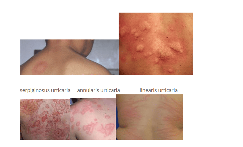
Acute urticaria may be caused by
- an IgE-mediated reaction,
- a process induced by a chemical substance (non-IgE urticaria),
- autoimmune urticaria
- cholinergic urticaria,
- cold urticaria
- mastocytosis (mast cell disease)
- periodic fever syndromes (e.g. Muckle-Wells34 syndrome)
- other causes
The release of the following bioactive substances may play a role in the development of acute urticaria:
- histamine,
- bradykinin,
- leukotriene C4,
- PGD2,
- other vasoactive substances released from mast cells and basophil leukocytes.
In terms of pathological processes,
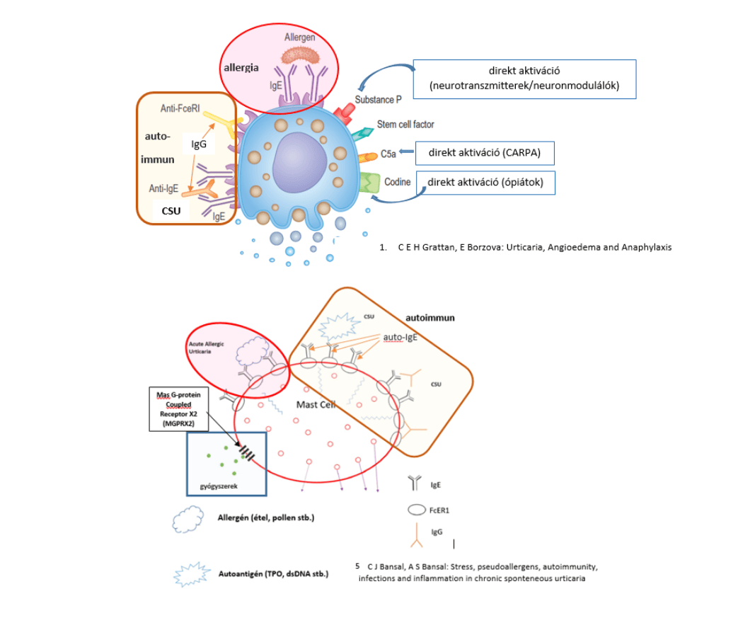
- in addition to type I hypersensitivity (allergy),
- type II antibody-mediated conditions involving cytotoxic T cell and/or complement activation and fibrin deposition lead to urticarial vasculitis, while
- type III hypersensitivity, characterised by immune complex formation, underlies the urticaria associated with SLE and other autoimmune diseases,
- certain medicines (e.g. opiates, vecuronium, succinylcholine, vancomycin, etc.) can cause mast cell degranulation directly or through other non-IgE mechanisms (see the previous summary, “Hypersensitivity, idiosyncrasy, pseudoallergy”),
- urticarias triggered by a physical stimulus include pressure urticaria, cold-induced urticaria and cholinergic urticaria (the latter may be provoked by emotional stress, heat or physical exertion).
In most cases of chronic spontaneous urticaria (CSU), no trigger can be identified (idiopathic urticaria), but a positive autologous serum skin test (ASST) indicates chronic autoimmune urticaria, and a special double-blind challenge (DBCT) can demonstrate a pseudoallergic background1-3,5.
Figure 2: Pathways of mast cell activation (based on references 1 and 5)
Urticaria may indicate a life-threatening process3 if
- angioedema appears on the lips, tongue or larynx,
- the urticarial lesions are painful, persist in one place for a long time (longer than 36–38 hours), are surrounded by bleeding, or leave pigmentation or bleeding after they fade (urticarial vasculitis is suspected, which can be confirmed by histological examination),
- it is accompanied by systemic symptoms (fever, joint and/or bone pain, lymphadenopathy),
- symptoms suggesting autoimmune thyroid disease or signs of another autoimmune disease are also present,
- liver enlargement, yellow sclera or pain below the right costal margin are noted,
- pneumonia or bronchospasm
- signs of fungal or bacterial skin infection are visible.
Skin symptoms resembling urticaria are seen in Schnitzler syndrome36: in addition to pink to red macules and slightly raised plaques on the skin (neutrophilic urticarial dermatosis), a monoclonal IgM gammopathy can be detected, accompanied by at least two of the following symptoms:
- fever,
- joint and/or bone pain,
- enlarged lymph nodes, spleen and/or liver,
- raised ESR,
- raised neutrophil leukocyte count,
- bone abnormalities on imaging.
It is also characterised by severe inflammation-related anaemia and amyloid A (AA) amyloidosis.
Angioedema is a pronounced, sudden, non-symmetrical (non-pitting) swelling of the subcutaneous tissues, which may occur with or without urticaria. Typical sites are the face, lips, tongue, larynx, genitals and limbs (although it may develop elsewhere too)6-10.
Figure: Angioedema
3.a with urticaria
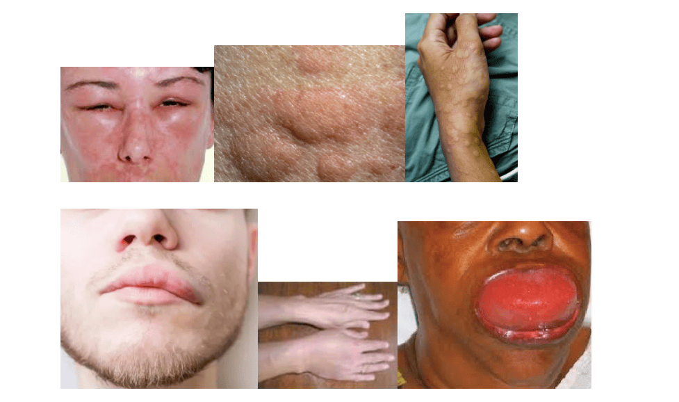
Angioedema (AE) not infrequently leads to a life-threatening condition. Whereas AE accompanied by urticaria is a (histaminergic) process caused by immune and non-immune mast cell activation, the forms without urticaria are mainly due to excessive activation of the kallikrein-kinin system, although in some cases the form without urticaria may also have a histaminergic background1,2,4,6-10.
Components of the kallikrein-kinin system:
- proteins [high- and low-molecular-weight (HMW&LMW) kininogens]
- enzymes (plasma and tissue kallikreins and their precursors: pre- and prokallikrein, as well as kininase I and the inactivating kininases)
- peptides (kinins: bradykinin, kallidin, des-Arg-bradykinin and des-Arg-kallidin)
- kinin receptors (B1 and B2 receptors)
Under the action of activated factor XII (FXIIa), prekallikrein is converted into the active enzyme plasma kallikrein, which cleaves the 9-amino-acid bradykinin from HMW kininogen (HK). Tissue kallikrein produces the 10-amino-acid kallidin from LMWK (or LK). The two kinins differ by one amino acid (kallidin has an additional N-terminal lysine). Under the action of an aminopeptidase (kininase I), kallidin is converted into bradykinin by removal of the lysine. This peptide acts on the B2 receptor. From both kinins, kininase I produces des-arginine peptides that act on the B1 receptor. Various kininases (kininase II) are involved in inactivating the active kinin peptides. Angiotensin-converting enzyme (ACE) also has this effect.
In addition to the kinin-inactivating kininases, the C1 esterase inhibitor (C1INH), which controls the classical and lectin pathways of the complement system, also plays a major role in maintaining the balance of the kallikrein-kinin system (by inhibiting the formation and activity of plasma kallikrein – see Figure 4).
In the congenital (hereditary) forms of angioedema (HAE), either an insufficient amount of C1INH is produced (type 1 HAE), or C1INH does not function properly (type 2 HAE), or continuously produced XIIa, resulting from an activating mutation of the Hageman factor gene, “keeps switched on” kallikrein-kinin activation. Less commonly, an altered endothelial surface due to angiopoietin-1 haploinsufficiency26 or the angiopoietin-1 gene (ANGPT1)27 activates the Hageman factor.
C1INH deficiency may also be acquired (acquired angioedema, AAE)1,2,4,31,32, due to autoimmune processes (e.g. IgG produced against endothelial cells in Gleich syndrome35) and malignant diseases1,2,4,7,28 (differentiation of HAE and AAE: Table 1.)
In certain individuals, ACE inhibitor (ACE-I) therapy used to control blood pressure can cause severe angioedema. This is because blocking one of the enzymes of the kininase II group that inactivate active kinins (ACE) leads to an accumulation of kinins and can cause angioedema and anaphylaxis32.
Figure 4: How the kallikrein-kinin system works and its link with the development of angioedema (AE)11-22
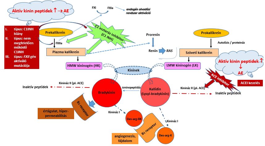
Table 1: Differentiation of hereditary and acquired angioedema on the basis of laboratory findings4
| C1INH level | C1INH activity | C1q level | C4 level | |
|---|---|---|---|---|
| Type 1 HAE | ¯ | ¯ | norm. | ¯ |
| Type 2 HAE | norm. | ¯ | norm. | ¯ |
| AAE | ¯ | ¯ | ¯ | ¯ |
| AE due to other causes (non-allergic, idiopathic) | norm. | norm. | norm. | norm. |
Anaphylaxis is a sudden-onset, potentially life-threatening condition whose main symptoms are bronchospasm and cardiovascular collapse, accompanied by urticaria, angioedema and gastrointestinal symptoms (vomiting, diarrhoea)1,2,4,6-10. Warning signs: reddening, burning and itching of the palms and soles, anxiety, fear of death, disorientation. Symptoms most often start on the mucous membranes, followed by respiratory, gastrointestinal and then cardiovascular signs (tachycardia, a drop in blood pressure, then bradycardia), and finally respiratory and circulatory failure.
The incidence of the condition in the EU and the US is 50–103 cases per 100,000 people per year, and mortality from anaphylaxis is 3.3 per 10,000,000 inhabitants1.
Although anaphylaxis may have immune (IgE and non-IgE) and non-immune (COX inhibition, direct mast cell activation, increased activity of the kallikrein-kinin system) causes, there is no significant difference in its course and symptoms. The common consequence of processes of different aetiology is the release of numerous bioactive substances, which leads to bronchospasm, increased mucus secretion, impaired myocardial contraction, increased vascular permeability, constriction of the peripheral and coronary arteries and dilatation of the small veins.
The most common triggers are foods, medicines, insect stings and latex. Less often, vaccines or inhaled aeroallergens are the activating factor. Anaphylaxis develops during their lifetime in 20% of patients with systemic mastocytosis (mast cell disease). In 60% of anaphylaxis cases presenting in outpatient settings and 20% of paediatric cases, no trigger can be identified. A multifactorial background is being confirmed in more and more cases. Cofactors lower the anaphylaxis threshold in about 30% of adult cases; the risk is higher among people taking β-blockers or ACE inhibitors1,2,4,32.
The incidence of food-induced anaphylaxis is 0.14 per 100,000 people per year, but in young children aged 0–4 years it may be as high as 7% per year1. The leading symptoms are usually bronchospasm and hypoxia. Food as a trigger most often appears in the first 10 years of life, but it is most severe in the teenage and young adult years. Although any food can cause anaphylaxis, peanuts, tree nuts, fish and shellfish do so most often. Rarely, anaphylaxis is triggered by physical exertion following food intake (exercise-induced anaphylaxis, EIA – e.g. WDEIA – wheat-dependent exercise-induced anaphylaxis – with production of anti-ω-gliadin IgE). An unusual form is α-gal (galactose-α-1,3-galactose) anaphylaxis, which appears not immediately but several hours after eating mammalian meat – in patients who have had a tick bite2.
Drug-induced anaphylaxis mostly occurs in hospital inpatients, with varying prevalence: it develops in 5–15 per 100,000 hospitalised patients after administration of an analgesic or antibiotic, while for dextran, penicillin, pentoxifylline and streptokinase this figure exceeds 30 per 100,000. Medicines are the leading cause of fatal anaphylaxis (1 in 50,000–100,000 cases), with parenteral administration posing the greatest risk.
In perioperative anaphylaxis (1 in 13,000 anaesthetics), the main triggers are neuromuscular blocking agents, antibiotics, blood and blood products, chlorhexidine and latex. Fatal cases occur more often in men, in people with cardiovascular disease and in those taking β-blockers.
Insect sting anaphylaxis can cause a life-threatening reaction in 0.4–0.8% of children and 3% of adults; 100 fatal cases are recorded in Europe each year. The leading symptoms are cardiovascular reactions. The severity of the reaction depends on the insect species, the amount of venom injected, the site of the sting, and the patient's age and existing conditions. In Hymenoptera venom allergy, allergen-specific immunotherapy provides protection against anaphylaxis after 8 weeks, for 3–5 years. In patients with mastocytosis, a fatal outcome may occur even at the first sting, especially if the venom contains certain components (e.g. phospholipase A2, a mast cell liberator).
In clonal mast cell disease (systemic mastocytosis or monoclonal mast cell activation syndrome), anaphylaxis may be triggered by Hymenoptera stings or medicines (NSAIDs, opiates, perioperative agents). If a patient has had unexplained anaphylaxis, systemic mastocytosis and monoclonal mast cell activation syndrome must be ruled out.
Rare causes of anaphylaxis: it should be considered when IgA-deficient patients are treated with transfusions or blood products, because in about a third of such patients circulating anti-IgA antibodies may trigger a life-threatening anaphylactic reaction to the IgA-containing blood product. Seminal fluid-induced anaphylaxis may occur in young atopic women and can be life-threatening in 20% of cases. Rupture of an Echinococcus hydatid cyst (during surgery or due to trauma) can also lead to anaphylaxis.
Four clinical forms have been described: immediate, biphasic, protracted and delayed. Symptoms may begin within seconds of encountering the trigger. The shorter the time between exposure and the onset of symptoms, the more severe the anaphylaxis. In biphasic cases, the attack recurs after 1–72 hours, but usually within 8–10 hours. This can be expected when the trigger is unknown and hypotension is present. Protracted anaphylaxis lasts longer than 24 hours and is often resistant to treatment. Late-onset anaphylaxis is extremely rare. The most severe cases involve laryngeal oedema, respiratory failure and circulatory collapse.
References
- C E H Grattan, E Borzova: Urticaria, Angioedema and Anaphylaxis 2019 Clinical Immunology 5th Edition
DOI: 10.1016/B978-0-7020-6896-6.00042-9 - J Pier, T A Bingemann: Urticaria, Angioedema, and Anaphylaxis, Pediatrics in Review 2020, 41(6): 283-292
Chapter 42: 585-600, https://doi.org/10.1542/pir.2019-0056 - H K Wong: Urticaria, emedicine, 2020, https://emedicine.medscape.com/article/762917
- C E Ciaccio: Angioedema: An Overview and Update, Missouri Medicine 2011, 108(5): 354-357,
https://www.ncbi.nlm.nih.gov/pmc/articles/PMC6188378/ - C J Bansal, A S Bansal: Stress, pseudoallergerns, autoimmunity, infections and inflammation in chronic sponteneous urticaria
Allergy, Asthma Clin Immunol 2019 15:56, https://doi.org/10.1186/s13223-019-0372-z - C Scully: Angioedema in Oral and Maxillofacial Medicine (3rd edition) 2013, p220-222,
https://doi.org/10.1016/B978-0-7020-4948-4.00032-5 - H H Lee: Agioedema, Medscape 2018, https://emedicine.medscape.com/article/135208
- M H Ohn&R Wadhwa: Angioneurotic Edema, In: StatPearls [Internet]. Treasure Island (FL): StatPearls Publishing;
2021. Jan, https://www.ncbi.nlm.nih.gov/books/NBK560611/ - B Zurav: An overview of angioedema: Pathogenesis and causes; UpToDate 2021
https://www.uptodate.com/contents/an-overview-of-angioedema-pathogenesis-and-causes - B Zurav: An overview of angioedema: Clinical features, diagnosis, and management; UpToDate 2021
https://www.uptodate.com/contents/an-overview-of-angioedema-clinical-features-diagnosis-and-management - Ch Golias, A Charalabopuolos et al.: The kinin system – bradykinin: biological effects and clinical implications. Multiple role of
the kinin system – bradykinin; Hippokratia 2007, 11(3):124-128; https://www.ncbi.nlm.nih.gov/pmc/articles/PMC2658795/ - H. Heiwald and W Müller-Esterl: Kinins in: S Offermans and W Rosentahl: Encyclopedic Reference of Molecular Pharmacology,
Springer-Verlag 2004 (ISBN 3-540-42843-7), p541-546 - Bhoola, K. D., & Fink, E. (2006). KALLIKREIN–KININ CASCADE in Encyclopedia of Respiratory Medicine, 483–493.
doi:10.1016/b0-12-370879-6/00204-0 - D J Campbell: Bradykinin and its related peptides in Handbook of Biologically Active Peptides 2006, p1175-1179
https://doi.org/10.1016/B978-012369442-3/50164-1 - D Regoli, F Marceau: Bradykinin Receptors in: J Enna& D B Bylund: xPharm: The Comprehensive Pharmacology Reference,
Esevier 2008 (ISBN 978-0-08-055232-3), p 1-4, 10.1016/B978-008055232-3.60203-X - Kenne, E., Rasmuson, J., Renné, T., Vieira et al.:. (2018). Neutrophils engage the kallikrein-kinin system to open up
the endothelial barrier in acute inflammation. The FASEB Journal, doi:10.1096/fj.201801329r - Y Pirahanchi, s Sharma: Physiology, Bradykinin In: StatPearls [Internet]. Treasure Island (FL): StatPearls Publishing; 2021 Jan.,
https://www.ncbi.nlm.nih.gov/books/NBK537187/ - I Isordia-Salas, R A Pixley et al.: The Role of Plasma High Molecular Weight Kininogen in Experimental Intestinal and Systemic
Inflammation, Archives of Medical Research 2005, 36:87–95; doi: 10.1016/j.arcmed.2005.02.001 - M K S Wong: Bradykinin in Handbook of Hormones 2016, Subchapter 30C 274-276
- K Majunder & J Wu: Molecular Targets of Antihypertensive Peptides: Understanding the Mechanisms of Action Based on the
Pathophysiology of Hypertension, Int. J. Mol. Sci. 2015, 16:256-283; doi:10.3390/ijms16010256 - A H Schmaier: The plasma kallikrein-kinin system counterbalances the renin-angiotensin system
J Clin Invest. 2002;109(8):1007–1009. https://doi.org/10.1172/JCI15490. - R Kahn, D Karpman: Kinin system activation in vasculitis, Acta Paediatrica 2011, 100:950-957,
DOI:10.1111/j.1651-2227.2011.02266.x - B Zuraw, H Farkas: Hereditary angioedema (due to C1 inhibitor deficiency): Pathogenesis and diagnosis, UpToDate
uptodate.com/contents/hereditary-angioedema-due-to-c1-inhibitor-deficiency-pathogenesis-and-diagnosis?search=hereditary
%20angioedema&source=search_result&selectedTitle=1~150&usage_type=default&display_rank=1 - B Zuraw, H Farkas: Hereditary angioedema: Epidemiology, clinical manifestations, exacerbating factors, and prognosis
https://www.uptodate.com/contents/hereditary-angioedema-epidemiology-clinical-manifestations-exacerbating-factors-and-
prognosis?search=hereditary%20angioedema&source=search_result&selectedTitle=3~150&usage_type=default&display_rank=3 - B Zuraw, H Farkas: Hereditary angioedema (due to C1 inhibitor deficiency): General care and long-term prophylaxis
https://www.uptodate.com/contents/hereditary-angioedema-due-to-c1-inhibitor-deficiency-general-care-and-long-term-
prophylaxis?search=hereditary%20angioedema&source=search_result&selectedTitle=5~150&usage_type=default&display_rank=5 - M d’Apolito, R Santacroce et al: Angiopoietin‐1 haploinsufficiency affects the endothelial
barrier and causes hereditary
angioedema, Clin Exp Allergy
. 2019 May;49(5):626-635. doi: 10.1111/cea.13349. - V Bafunno, D Firinu et al.: Mutation of the angiopoietin-1 gene (ANGPT1) associates with a new type of hereditary angioedema
J Allergy Clin Immunol 2018, 141(3):1009-1017, http://dx.doi.org/10.1016/j.jaci.2017.05.020 - M. Maurer, M Magerl et al.: The international WAO/EAACI guideline for the management of hereditary angioedema—
The 2017 revision and update, Allergy 2018, 73(8):1575-1596, https://doi.org/10.1111/all.13384 - P J Busse, S C Christiansen et al.: US HAEA Medical Advisory Board 2020 Guidelines for the Management of Hereditary
Angioedema, J Allergy Clin Immunol Pract 2021, 9(1):132-50, https://doi.org/10.1016/j.jaip.2020.08.046 - S Saini: Acquired C1 inhibitor deficiency: Clinical manifestations, epidemiology, pathogenesis, and diagnosis, UpToDate
https://www.uptodate.com/contents/acquired-c1-inhibitor-deficiency-clinical-manifestations-epidemiology-pathogenesis-and-
diagnosis?search=Acquired%20C1%20inhibitor%20deficiency&source=search_result&selectedTitle=1~32&usage_type=default
&display_rank=1 - S Saini: Acquired C1 inhibitor deficiency: Management and prognosis, UpToDate
https://www.uptodate.com/contents/acquired-c1-inhibitor-deficiency-management-and-prognosis?search=Acquired%20C1
%20inhibitor%20deficiency&source=search_result&selectedTitle=2~32&usage_type=default&display_rank=2 - A C Guyer, A Banerji: ACE inhibitor-induced angioedema, UpToDate
https://www.uptodate.com/contents/ace-inhibitor-induced-angioedema?search=ace%20inhibitor
%20induced%20angioedema&source=search_result&selectedTitle=1~150&usage_type=default&display_rank=1 - D J Pearson: Psychologic and somatic interrelationships in allergy and pseudoallergy, J Allergy Clin Immunol 1986,
81(2):351-360, DOI: 10.1016/0091-6749(88)90900-1 - R A Schwartz: Wells syndrome 2021 March, emedicine, https://emedicine.medscape.com/article/1124844-overview
- R Haaber, J A Chebi et al.: Gleich syndrome, International Journal of Dermatology 2020, https://doi.org/10.1111/ijd.14963
- D Lipsker: Schnitzler syndrome, Orphanet Journal of Rare Diseases 2010,5:38, http://www.ojrd.com/content/5/1/38

SIBO and food intolerances: is food necessarily to blame?
Bloating after milk, bread or onions? The way the digestive system works may also play a role behind these symptoms. Dr Anikó Hovanecz’s article expla...

A corporate health programme in practice – the SFC CEO’s experience
What does caring for employees and their family members mean for a company leader? Through his personal experience, Gábor Horváth, CEO of SFC, explain...

Don't just get the school bag ready
Starting school is not only about new exercise books and a changed daily routine but also about reorganising family meals. The FOODTEST Children's pac...