In this article
Author: Dr Károly Krkos
In the last century, the frequency of autoimmune diseases overtook that of infectious diseases in developed countries. Genetic and environmental factors and the state of the microbiome play a role in the development of these multifactorial diseases. The treatments currently available for autoimmune conditions are based on weakening the immune system and increase the risk of malignant tumours and infections. In recent decades it has become clear that these conditions are characterised by the development of a different, “dysbiotic” microbiome, and that the effectiveness of treatment in patients with inflammatory bowel diseases (IBD) or rheumatoid arthritis is also influenced by the composition of the gut microbiome and the metabolism of the drug used. The microbiome modifies innate and adaptive immunity directly or by transforming dietary metabolites, so changing the composition and activity of the microbiome may contribute to the prevention and treatment of autoimmune diseases.
Figure (1): The interaction between diet and the microbiome, and host health (based on reference 32)
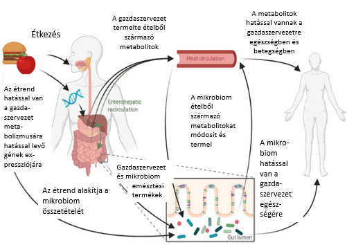
The host and the microbiota interact with each other in a complex process that depends on the host’s genes and immune system as well as on the composition and function of the gut microbiota. The host’s gene expression is influenced by diet. Metabolites are produced from food during digestion. Products that reach the colon have a direct effect on the composition of gut microbes. The microbes further transform the products made by the body into microbiome metabolites. Both host and microbiome metabolites enter the circulation through enterohepatic recirculation or absorption across the intestinal epithelium.
Despite the large differences in dietary patterns, the microbiome of people living with meat and processed food consumption has undergone significant change, such as the development of an abundance of microbial carbohydrate-active enzymes (CAZymes)6,7. CAZymes play a significant role in breaking down carbohydrates and in modifying the synthesis of glycoconjugates, oligosaccharides and polysaccharides. Besides dietary changes, these processes were also influenced by other factors (e.g. the introduction of antibiotics in the treatment of diseases). The recognition that dietary changes have contributed significantly to the rising frequency of autoimmune diseases led to the development of a special dietary protocol (the autoimmune protocol, AIP), the essence of which is to exclude from the diet foods that may be able to trigger inflammatory processes in the gut. The Western-type diet does not support the development of a diverse, resilient microbiota. According to the hygiene hypothesis, a hyper-hygienic environment early in life does not help normal immune tolerance to develop. Recent literature supports the view that when autoimmune diseases appear, the barrier function of the gut is impaired, and the entry and detection of atypical external antigens thus leads to immune dysregulation2,3,4,5.
The gut microbiome and host immunity
Early in life, the interaction between microbial colonisation and the host’s immune system induces a pro-inflammatory or anti-inflammatory predominance that has lifelong effects. Caesarean section, not breastfeeding and antibiotic use can disrupt these processes. Oligosaccharides in breast milk promote colonisation by Bifidobacterium species, which become the predominant component of the early microbiota and produce fucose, acetate, pyruvate and 1,2-propanediol, thereby promoting microbiota expansion and helping tolerance to the commensal flora to develop8,9. After solid food is introduced (at around 6 months of age), the bacterial community changes significantly, producing large amounts of various metabolites (e.g. butyrate, which promotes the maturation of the colonic mucosa10 and inhibits the growth of bacteria with high pathogenic potential11). Inappropriate TLR stimulation caused by microbial antigens may affect the progression of autoimmune disease (TLR7 overexpression is observed in a mouse model of SLE11). In this experiment, lupus progression could be reduced by feeding resistant starch, thereby changing the composition of the microbial flora and metabolites11. In an animal study of multiple sclerosis, stimulating the TLR2 receptor with bacterial lipoproteins reduced disease severity12. These results support the view that the molecular patterns characteristic of gut microorganisms are important regulators of TLR signalling, contributing to a tolerogenic gut environment12.
Studies in animal models have demonstrated the development of autoimmune diseases based on altered microbiome patterns and dysfunction of the gut barrier2,13,14. Dietary changes affect the composition and metabolite production of the microbiome. Gut microbes act on innate and adaptive immunity directly (through microbe-associated molecular patterns – MAMPs – and pathogen-associated molecular patterns – PAMPs) or indirectly (through changes in metabolite production). A gut barrier damaged by an injured mucus layer or weakened tight junctions contributes to atypical antigen detection by antigen-presenting cells (dendritic cells, macrophages). The gut microbiome, which can modify toll-like receptor (TLR) tolerance15, may also play a role in exacerbating autoimmune diseases, since inadequate TLR activation triggered by commensal bacteria can disrupt immune regulation16,17. In autoimmune patients, the CD4+ T-cell population shifts towards disease-activating Th1 and Th17 cells, while the proportion of regulatory T cells (Treg) decreases. All this affects the production of microbial metabolites (such as butyrate18 or propionate19).
Microbiome-induced adaptive immune dysregulation is observed in autoimmune patients. The microbiome of patients with multiple sclerosis contains higher proportions of Akkermansia muciniphila and Acinetobacter calcoaceticus, whose role in gut homeostasis is uncertain; in vitro they stimulate Th1 differentiation but not differentiation towards Treg17. The commensal flora of the mammalian gut, mainly Clostridium clusters IV and XIVa, which can break down dietary fibre into butyrate, induce Treg development, and in a mouse model their abundance is inversely proportional to the severity of colitis18. Characteristically, in autoimmune diseases the Treg/effector T ratio shifts away from the homeostatic level: the number of tolerogenic, IL-10-producing CD25+FOXP3+ T (Treg) cells decreases relative to effector Th1 and Th17 cells20. These changes may contribute to increased intestinal permeability because of the pro-inflammatory cytokines produced (TNFα, IL-17), since these modify tight junction proteins – this has been shown in mouse models21,24 and in patients with multiple sclerosis22,23.
Diet and the microbiome
Changing the diet can modify the composition of the microbiome and of the metabolites it produces25-28. A marked increase in Bacteroides strains relative to Prevotella is accompanied by a decrease in genes for fibre-degrading enzymes29.
Numerous randomised controlled studies have been carried out on dietary carbohydrate fermentation, evaluating the effect of fibre intake on microbiome composition or activity. Microbiome changes differ between individuals, but the response to a selected fibre structure is surprisingly consistent34. For example, a dose-response study of maize resistant starch (RS4) observed an increase in Eubacterium rectale, whereas tapioca RS4 caused an increase in Parabacteroides distasonis33.
By definition (International Scientific Association for Probiotics and Prebiotics), prebiotics and postbiotics are substrates selectively used by host microorganisms that have beneficial health effects35. In a study of a high-fibre diet (a 17-week prospective, randomised study), changes in participants’ CAZyme profiles were observed without any change in SCFA production36. The amount of fibre taken in correlated with the carbohydrate content of the stool, suggesting that the microbiota was unable to break down the fibre completely because the fibre-degrading pathway had become saturated. Increasing fibre intake does not directly mean increased SCFA synthesis; this also depends on the state of the microbiota. A study analysing a group of patients with ulcerative colitis found that the main butyrate-producing bacteria – Roseburia hominis and Faecalibacterium prausnitzii – were reduced in those affected compared with healthy people, and the extent of the reduction varied in parallel with disease activity37,38. On this basis, the level of bacterial SCFA production is a better indicator than the amount of fibre taken in. SCFA production is necessary for immune homeostasis and for healthy gut barrier function39. This drew attention to the beneficial effect on host health of metabolites produced by microorganisms, the postbiotics. Animal and human studies have shown that inadequate butyrate production causes β-oxidation in mucus-producing goblet cells to be replaced by anaerobic glycolysis, which has a negative effect on goblet cell differentiation and on the production of MUC2 (the main glycoprotein component of mucus)40. This increases the microbes’ exposure to oxygen41, which has an unfavourable effect on the main butyrate-producing anaerobes42. Another beneficial effect of butyrate is that it downregulates the transcription of pro-inflammatory cytokines and promotes the differentiation of Th cells into Treg cells39,43. The other main SCFA, propionate, plays an important role in hepatic gluconeogenesis, in the development of satiety and in lowering cholesterol44. In a group of patients with multiple sclerosis, supplementing routine treatment with propionate achieved remission, and the number of Treg cells increased while the numbers of Th1 and Th17 cells decreased45.
Exclusive or elimination diets aim to optimise SCFA production. In IBD patients this is not simple, as patients complain of bloating and excessive gas after eating fibre46. Although complex carbohydrates are not recommended during the active phase of the disease or after surgery, they may play a role in the diet during remission. Exclusive (EEN) or partial enteral nutrition (PEN) is recommended for IBD patients. In the former case, this may be an elemental (containing amino acids), semi-elemental (containing oligopeptides) or polymeric (containing whole proteins) liquid diet. In PEN, the liquid nutrition is supplemented with solid food based on the CDED – the Crohn’s disease exclusion diet. Its essence is that it is rich in energy and protein, low in fat, free of coarse fibre and dairy, and contains no simple sugars. The CDED reduces the abundance of Proteobacteria and inflammation. EEN supports remission in Crohn’s disease and changes the metabolite production of the colonic microbiota: protein fermentation dominates over carbohydrate fermentation47. It is not clear what factor actually induces remission: the lack of fibre, the lack of other dietary components, the reduction in bacterial diversity and density, or a combination of all of these1.
Proteins are broken down during digestion by the host’s enzymes into amino acids and smaller peptides, most of which are absorbed from the small intestine. Some of the peptides may reach the colon, where they are transformed by microbes. Based on experimental data, 17–38% of these are converted into short-chain fatty acids (SCFAs). During protein degradation, the commensal flora also produces essential amino acids and branched-chain fatty acids (BCFAs), which serve as an alternative energy source for colonocytes49. Toxic substances are also produced: ammonia, hydrogen sulphide and phenols, which have been shown in animal experiments (mice) to increase intestinal permeability48. In a mouse model of IBD, feeding with animal proteins caused colitis to flare up and pro-inflammatory macrophages to be activated, whereas plant proteins did not trigger such processes50. Apart from the fact that every amino acid is deaminated with the formation of ammonia, different proteins affect bacteria differently depending on their biochemical characteristics:
- BCFAs (branched-chain fatty acids) are formed only from branched-chain amino acids51;
- sulphated amino acids favour sulphate-reducing, hydrogen sulphide-producing bacteria and cause their expansion, as has been observed in patients with ulcerative colitis52;
- aromatic amino acids are favoured mainly by Enterobacter and Escherichia species, which form phenols from them;
- the products formed when one of the essential amino acids, tryptophan, is metabolised by microbes (the indole pathway) bind to the host’s aryl hydrocarbon receptors, reduce the expression of pro-inflammatory cytokines and chemokines and promote the production of IL-22, which prompts epithelial cells to release antimicrobial peptides and regulates the growth of the microbiota53,54;
- tryptophan intake or tryptophan-producing Lactobacillus strains alleviated colitis in mouse experiments53-55.
Besides being a nutrient source for gut microbes, dietary fat affects the composition of the microbiota through increased bile acid production56,57 and promotes the growth of commensal bacteria with high pathogenic potential58 (e.g. Bilophila wadsworthia). Changes in the composition of the microbiota due to fat intake may alter the amount of secondary bile acids produced by microbes. High-fat food is associated with an increase in lipopolysaccharides (LPS) and induction of the pro-inflammatory TLR4 pathway, leading to low-grade inflammation that has a fundamental effect on autoimmune activity60,61. Similarly, an increase in Gram-negative bacteria represents a source of LPS and a pro-inflammatory effect60, which Bifidobacterium species, able to reduce LPS-induced inflammation, could counterbalance – so a decrease in these also favours pro-inflammatory processes60. On the other hand, eating food rich in unsaturated fatty acids (e.g. walnuts) enriches Faecalibacterium, Roseburia and Clostridium strains, which reduce the production of pro-inflammatory secondary bile acids derived from microbes59.
Food processing includes the following: heat (e.g. cooking), mechanical action (e.g. slicing, grinding, whipping) and the use of additives, to increase shelf life and digestibility and to improve the taste and appearance of food. Eating raw tubers rich in starch reduces the amount of bacteria and increases microbiome metabolism compared with the microbiota of people eating cooked tubers62. Microbial metabolism may be beneficial or toxic for the host and may also modify the effect of medicines63. Treating wholemeal wheat flour with cooking–freezing cycles significantly increases the proportion of resistant starch, whose in vitro fermentation raises propionate levels and the number of Bifidobacteria64. As the use of additives has become increasingly common, autoimmune diseases have multiplied, and damage to tight junction integrity and increased intestinal permeability may play a role in this65. A general principle in applying the various (exclusion, elimination) diets is the removal of industrially processed food products and foods containing additives from the diet.
Options for modifying the microbiota
Microbial diversity is low in patients with various autoimmune diseases67, and enriching diversity benefits the microbiota and thus the patient66. However, it cannot be said that a highly diverse microbiome is the same as a healthy microbiome. Machine learning is an effective tool for making use of existing metagenomic and metabolomic data to better understand the links between autoimmune diseases and the microbiome68.
(Machine learning: a computer-based method in which machines are taught how to use data more effectively. The machines become able to learn without being explicitly programmed70.)
The host’s genetic factors and the state of their existing microbiota (among other factors) can influence how the patient responds to a change in diet26,69,71-73. More and more knowledge is accumulating about personalised changes to gut microbial composition with the help of diet.
For an optimal intervention, the state of the disease, the biomarkers characteristic of the disease, the composition and function of the gut microbiome, and the drug treatment in place must be taken into account. The therapeutic diet must be set individually, preceded by predictive modelling: human randomised controlled trials and cross-over dietary interventions are needed to ensure remission of the disease, and the results of animal studies must be evaluated in human studies, with the experience gained there fed back into experimental work. [For example, a human microbiome-associated (HMA) rodent model is colonised with patient and control samples, and these animals are studied on diets of different composition.]1
Influencing the gut microbiota
There are numerous options for manipulating the gut microbial system with the aim of restoring a healthy microbiota. These techniques are:
- faecal microbiome transplantation (FMT) and its combination with special diets or dietary supplements;
- orthogonal niche engineering.
Combined use of FMT and diet can effectively improve the patient’s condition, although its mechanism of action has not yet been fully explored. The procedure was first used, successfully, to treat recurrent gastrointestinal Clostridioides difficile infection (CDI): about 90% of patients became symptom-free with the method75. The effect of FMT treatment has also begun to be studied in many other diseases: T1DM76, multiple sclerosis77, coeliac disease78, rheumatoid arthritis79 – for these, few data are available so far. Ulcerative colitis80-83, Crohn’s disease84-85, liver diseases86,87, metabolic syndrome88 and pathological neurological processes89 are better studied conditions, but the effectiveness of treatment in these conditions has not come close to the success of CDI treatment. The hypothesis that a certain microbiota composition can trigger the disease or worsen the patient’s condition (e.g. in certain subgroups of IBD90) can still be said to hold, even though the imperfect results are well documented owing to certain pitfalls and causes91-93. Although not thoroughly researched, two approaches are considered noteworthy:
- making the recipient’s diet similar to the donor’s, so that the effect of the donor microbiota is more reliable in the new host, or
- trying to strengthen the beneficial components of the healthy donor microbiota with the CDED diet or with inulin-type fructans, which support the growth of the SCFA-producing Roseburia and Faecalibacterium species74,96.
FMT alone is only partly successful in relieving symptoms94 or inducing remission80. In mice with emphysema, FMT supplemented with a high-fibre diet had a favourable effect on microbiota composition and SCFA production95. Where the composition of the donor microbiota is not known, designing a diet to support FMT is limited. The aim should be to use a well-defined culture derived from the stool of a healthy donor97,98.
Probiotic–diet combinations and synbiotics are an option independent of FMT, in which one or more microbial strains (e.g. VSL#399) are given as a probiotic. This is a frequently used therapeutic option in the treatment of IBD patients98,100,101. The matrix in which the probiotic is delivered may be a food (e.g. yoghurt, cereal bar) or a capsule containing a protective emulsion or biopolymer. Which form is chosen matters for preserving the viability of the bacteria taken in, since the selected microbes may be sensitive to changes in pH (e.g. the strongly acidic environment of the stomach) and to unsuitable oxygen levels (the commensal flora contains many obligate anaerobes). For successful colonisation, besides adequate protection of the microbes, it is also important that they have access to suitable nutrients102.
Prebiotics and probiotics can be combined; these are synbiotics. Complementary synbiotics consist of a beneficial but mutually independent prebiotic–probiotic mixture, whereas in synergistic synbiotics the prebiotic supports the function of the probiotic, improving its chances of survival and its integration into the host microbiota30. The study of synbiotics in humans has only just begun, but certain combinations have been found effective in treating autoimmune patients. Combining Bifidobacteria and Lactobacilli with psyllium husk104, fructo-oligosaccharides105 and inulin106,107 proved beneficial in IBD patients. Similarly, a mixture of Lactobacillus acidophilus, Lactobacillus casei and Bifidobacterium bifidum supplemented with inulin108 showed a beneficial effect in rheumatoid arthritis, as did Lactobacillus sporogenes combined with maltodextrin and fructo-oligosaccharides109 in type 1 diabetes.
Orthogonal niche engineering is a further development of the synbiotic concept in that it exploits the microorganism–substrate relationship to integrate bacteria into the host microbiota110,111.
[Niche (ecological niche): the set of factors, or their interaction, thanks to which certain species and populations are able to survive, i.e. they do not die out and are not at risk of extinction112.]
Whereas the prebiotics in synbiotics are generally everyday nutrients, orthogonal niche engineering uses components that are not used, or only rarely used, as prebiotics for this task. For example, porphyran, a sulphated polysaccharide derived from a seaweed (genus Porphyra), is used to introduce human commensal microbiota into the mouse gut110,111. The ability of the human microbiome to break down seaweed is more widespread than originally assumed. Orthogonal niche engineering allows the bacteria introduced to remain viable in the gut microbiome and their genes to be utilised113. Processes such as the breakdown of agarose, porphyran and carrageenan can be used to introduce beneficial bacteria that are either wild-type, modified versions of an original strain, or have been transfected with genes able to use the orthogonal nutrient. The method offers the possibility of reintroducing into the human gut flora bacteria that were lost during industrialisation. One example is Lactobacillus reiteri, whose immunomodulatory effect in SLE and multiple sclerosis has been studied in detail in mouse experiments114,115. Another direction is the creation of recombinant bacteria that produce therapeutic compounds. For example, Streptococcus thermophilus and Lactococcus lactis strains were transfected with the IL10 gene to treat animals with IBD116, and the human defensin-5 gene was introduced into the DNA of a Lactococcus lactis strain, which alleviated dextran sulphate-induced colitis in experimental animals by blocking the NF-kB pathway117.
Unusual nutrients allow the extent and duration of colonisation to be tailored to the individual. Prolonged, continuous intake of a specialised nutrient may, however, lead to reduced activity of the implanted bacteria110. A further problem may be that the special property (the gene with which the introduced bacterium was transfected) passes to other bacteria through horizontal gene transfer103, which may lead to reduced specificity or a completely altered response.
According to a meta-analysis of epidemiological studies, a higher fibre intake (25–29 g/day) would lead to a significant reduction in the incidence of non-communicable diseases (e.g. coronary heart disease, stroke, type 2 diabetes, colon cancer). Although promising data show the importance of dietary change and the state of the microbiome in autoimmune diseases, their mechanism of action is not known in every respect. For example, people whose gut flora contains a higher proportion of Prevotella copri at the expense of Bacteroides species are more prone to developing rheumatoid arthritis (RA)31,118. The severity of symptoms in RA patients can be reduced with various (vegan, gluten-free) diets119. This link is uncertain120, even though numerous studies support the effectiveness of microbiome and dietary change121.
Several challenges remain in the field of microbiome manipulation. Because of the resilience of the human microbiota, the results of animal experiments do not produce in human studies the pronounced change that the experiments showed122. Although dietary change can be an effective tool for altering the gut flora, stopping the diet may lead to a return to the microbiota’s previous state123. Many further studies and convincing results are needed before personalised microbiota manipulation can become part of routine treatment.
References
- M Wolter, E T Grant et al.: Leveraging diet to engineer the gut microbiome 2021, Nature Reviews Gastroenterology and Hepatology, 18(12):885-902, DOI: https://doi.org/10.1038/s41575-021-00512-7
- N J Wasko, F Nichols, R B Clark: Multiple sclerosis, the microbiome, TLR2, and the hygiene hypothesis 2020, Autoimmunity Reviews, 19(1):102430, https://doi.org/10.1016/j.autrev.2019.102430
- W E Ruff, T M Greiling, M A Kriegel: Host-microbiota interactions in immune-mediated diseases 2020, Nature Reviews Microbiology, 18:521-538, https://doi.org/10.1038/s41579-020-0367-2
- B Li, C Selmi et al.: The microbiome and autoimmunity: a paradigm from the gut-liver axis 2018, Cellular& Molecular Immunology, 15:595-609, https://doi.org/10.1038/cmi.2018.7
- A Márquez et al.: Meta-analysis of immunochip data of four autoimmune diseases reveal novel single-disease and cross-phenotype associations 2018, Genome Medicine, 10:97, https://doi.org/10.1186/s13073-018-0604-8
- A E Kaoutar, F Armougom et al: The abundance and variety of carbohydrate-active enzymes in human gut microbiota 2013, Nature Reviews Microbiology, 11:497-504, https://doi.org/10.1038/nrmicro3050
- S A Smits et al.: Seasonal cycling in the gut microbiome of the Hadza hunter-gatherers of Tanzania 2017, Science, 357:802-806, https://doi.org/10.1126/science.aan4834
- M A E Lawson et al: Breast milk-derived human milk oligosaccharides promote Bifidobacterium interactions within a single ecosystem. 2020, ISME J, 14:635-648, https://doi.org/10.1038/s41396-019-0553-2
- S Fanning et al.: Bifidobacterial surface-exopolysaccharide facilitates commensal-host interaction through immune modulation and pathogen protection 2012, PNAS, 109:2108-2113
- M Beaumont et al.: Gut microbiota derived metabolites contribute to intestinal barrier maturation at the suckling-to-weaning transition 2020, Gut, 11:1268-1286, https://doi.org/10.1080/19490976.2020.1747335
- D F Zegarra-Ruiz et al.: A diet-sensitive commensal lactobacillus strain mediates TLR7-dependent systemic autoimmunity 2019, Cell Host & Microbe, 25:113-127.e6, https://doi.org/10.1016/j.chom.2018.11.009
- M Fujiwara et al.: Enhanced TLR2 responses in multiple sclerosis 2018, Clinical Experimental Immunology, 193:313-326, https://doi.org/10.1111/cei.13150
- D Zheng, T Liwinski, E Elinav: Interaction between microbiota and immunity in health and disease 2020, Cell Research, 30:492-506, https://doi.org/10.1038/s41422-020-0332-7
- B Li, C Selmi et al.: The microbiome and autoimmunity: a paradigm from the gut-liver axis 2018, Cellular and Molecular Immunology, 15:595-609, https://doi.org/10.1038/cmi.2018.7
- E D’Hennezel, S Abubucker et al.: Total lipopolysaccharide from the human gut microbiome silences toll-like receptor signaling 2017, mSystems, 2:e00046-17, https://doi.org/10.1128/mSystems.00046-17
- D R Zegarra-Ruiz et al.: A diet-sensitive commensal Lactobacillus strain mediates TLR7-dependent systematic autoimmunity 2019, Cell Host & Microbe, 25:113-127.e6, https://doi.org/10.1016/j.chom.2018.11.009
- E Cekanaviciute et al.: Gut bacteria from multiple sclerosis patients modulate human T cells and exacerbate symptoms in mouse model 2017, PNAS, 114:10713-10718, https://doi.org/10.1073/pnas.1711235114
- K Atarashi et al.: Induction of colonic regulatory T cells by indigenous Clostridium species 2011, Science, 331(6015):337-341, https://doi.org/10.1126/science.1198469
- A Dusha et al.: Propionic acid shapes the multiple sclerosis course by an immunomodulatory mechanism 2020, Cell, 180:1067-1080.e16, https://doi.org/10.1016/j.cell.2020.02.035
- N Ohkura, Y Yasumizu et al.: Regulatory T-cell specific epigenomic region variants are a key determinant of susceptibility to common autoimmune diseases 2020, Immunity, 52:1119-1132.e4, https://doi.org/10.1016/j.immuni.2020.04.006
- J S Lee, C M Tato et al.: Interleukin-23-independent IL17 production regulates intestinal epithelial permeability 2015, Immunity, 43:727-738, https://doi.org/10.1016/j.immuni.2015.2015.09.003
- I Cosorich, G Dalla-Costa et al.: High frequency of intestinal Th17 cells correlates with microbiota alterations and disease activity in multiple sclerosis 2017, Science Advances, 3:e1700492, https://doi.org/10.11266sciadv.1700492
- M C Buscarinu, B Cerasoli et al.: Altered intestinal permeability in patients with remitting-relapsing multiple sclerosis: a pilot study 2017, Multiple Sclerosis Journal, 23:442-446, https://doi.org/10.1177/1352458516652498
- X Zhang, B D Chen et al.: The gut microbiota: emerging evidence in autoimmune diseases 2020, Trends in Molecular Medicine, 26:862-873, https://doi.org/10.1016/j.molmed.2020.04.001
- R N Carmodi, G K Gerber et al.: Diet dominates host genotype in shaping the murine gut microbiota 2015, Cell Host & Microbiome, 17:72-84, https://doi.org/10.1016/j.chom.2014.11.010
- A Wenkataraman, J R Sieber et al.: Variable responses of human microbiomes to dietary supplementation with resistance starch 2016, Microbiome, 4:33, https://doi.org/10.1186/s40168-016-0178-x
- G D Wu, C Compher et al.: Comparative metabolomics in vegans and omnivores reveal constraints on diet-dependent gut microbiota metabolite production 2016, Gut, 65-63-72, https://doi.org/10.1136/gutjnl-2014-308209
- A M Sheflin, C L Melby et al.: Linking dietary patterns with gut microbial composition and function 2017, Gut Microbes, 8:113-129, https://doi.org/10.1080/19490976.2016.1270809
- P Vangay, A J Johnson et al.: US immigration westernizes the human gut microbiome 2018, Cell, 175:962-972.e10, https://doi.org/1016/j.cell.2018.10.029
- K S Swanson et al: The International Scientific Association for Probiotics and Prebiotics (ISAPP) consensus statement on the definition and scope of synbiotics. 2020, Nature Reviews Gastroenterology and Hepatology, 17:687-701, https://doi.org/10.1038/s41575-020-0344-2
- J C Clemente, J Manasson, J U Scher: The role of gut microbiome in systematic inflammatory disease 2018, BMJ, 360:j5145, https://doi.org/10.1136/bmj.j5145
- A J Johnson, J J Zheng et al.: A guide to diet and microbiome study design 2020, Frontiers in Nutrition, 7:79. https://doi.org/10.3389/fnut.2020.00079
- E C Deehan, C Yang et al.: Precision microbiome modulation with discrete dietary fiber structures directs short-chain fatty acid production 2020, Cell Host & Microbiome, 27:389-404.e6, https://doi.org/10.1016/j.chom.2020.01.006
- D So, K Whelan et al.: Dietary fiber intervention on gut microbiota composition in healthy adults: a systematic review and meta-analysis 2018, American Journal of Clinical Nutrition, 107:965-983, https://doi.org/10.1093/ajcn/nqy041
- G R Gibson, R Hutkins et al.: Expert consensus document: the International Scientific Association for Probiotics and Prebiotics (ISAPP) consensus statement on the definition and scope of prebiotics 2017, Nature Reviews Gastroenterology and Hepatology, 14:491-502, https://doi.org/10.1038/nrgastro.2017.75
- H C Wastyk, G K Fragiadakis et al.: Gut microbiota-targeted diets modulate human immune status 2021, Cell, 184:4137-4153, https://doi.org/10.1016/j.cell.2021.06.019
- K Machiels, M Joossens et al.: A decrease of the butyrate-producing species Roseburia hominis and Faecalibacterium prausnitzii defines dysbiosis in patients with ulcerative colitis 2014, Gut, 63:1275-1283, https://doi.org/10.1136/gutjnl-2013-304883
- H Earley, G Lennon et al.: Colonization of the colonic mucus gel layer with butyrogenic and hydrogenotropic bacteria in health and ulcerative colitis 2021, Scientific Reports, 11:7262, https://doi.org/10.1038/s41598-021-86166-6
- D P Venegas, M K De la Fuente et al.: Short chain fatty acids (SCFAs) mediated gut epithelial and immune regulation and its relevance for inflammatory bowel diseases 2019, Frontier in Immunology, 10:277, https://doi.org/10.3389/fimmu.2019.00277
- A Sünderhauf, M Hicken et al.: Loss of mucosal p32/gC1qR/HABP1 triggers energy deficiency and impairs goblet cell differentiation in ulcerative colitis 2021, Cellular and Molecular Gastroenterology and Hepatology, 12:229-250, https://doi.org/10.1016/j.jcmgh.2021.01.017
- M X Byndloss, E E Olsan et el.: Microbiota activated PPAR-g signaling inhibits dysbiotic Entrobacteriaceae expansion. 2017, Science, 357:570-575, https://doi.org/10.1126/science.aam9949
- M Y Zheng, N Imohara and G Nuňez: Mechanisms of inflammation-driven bacterial dybiosis in the gut. 2017, Mucosal Immunology, 10:18-26, https://doi.org/10.1038/mi.2016.75
- Y Furusawa et al.: Commensal microbe-derived butyrate induces the differentiation of colonic regulatory T-cells. 2013, Nature, 504:446-450, https://doi.org/10.1038/nature12721
- P Luis, HJ Flint: Formation of propionate and butyrate by the human colonic microbiota. 2017, Environmental Microbiology, 19:29-41, https://doi.org/10.1111/1462-2920.13589
- A Duscha, B Gisevius et al.: Propionic acid shapes the multiple sclerosis disease course by an immunomodulatory mechanism. 2020, Cell, 180:1067-1080.e16, https://doi.org/10.1016/j.cell.2020.02.035
- A B Cohen, D Lee et al.: Dietary patterns and self-reported association of diet with symptoms of inflammatory bowel disease. 2013, Digestive Diseases and Sciences, 58:1322-1328, https://doi.org/10.1007/s10620-012-2373-3
- C Walton et al: Enteral feeding reduces metabolic activity of the intestinal microbiome in Crohn’s disease: an observational study. 2016, European Journal of Clinical Nutrition, 70:1052-1056, https://doi.org/10.1038/ejcn.2016.74
- S R Llewellin, G J Britton et al: Interaction between diet and the intestinal microbiota alter intestinal permeability and colitis severity in mice. 2018, Gastroenterology, 154:1037-1046e.2, https://doi.org/10.1053/j.gastro.2017.11.030
- G T Macfarlane, G R Gibson et al: Estimation of short-chain fatty acid production from protein by human intestinal bacteria based on branched-chain fatty acid measurements 1992, FEMS Microbiology Letters, 101:81-86, https://doi.org/10.1111/j.1574-6968.1992.tb05764.x
- K Kostovcikova, S Coufal et al: Diet rich in animal protein promotes pro-inflammatory macrophage response and exacerbates colitis in mice. 2019, Frontiers in Immunology, 10:919, https://doi.org/10.3389/fimmu.2019.00919
- K J Portune, M Beaumont et al: Gut microbiota role in dietary protein metabolism and health-related outcomes: the two sites of coin. 2016, Trends in Food Science and Technology, 57B:213-232, https://doi.org/10.1016/j.tifs.2016.08.011
- G R Gibbson, J H Cummings, G T Macfarlain: Growth and activities of sulfate-reducing bacteria in gut contents of healthy subjects and patients with ulcerative colitis. 1991, Trends in Food Science and Technology, 86:103-111, https://doi.org/10.1111/j.1574-6968.1991.tb04799.x
- B Lamas, M L Richard et al: CARD9 impacts colitis by altering gut microbiota metabolism of tryptophan into aryl hydrocarbon receptor ligands. 2016, Nature Medicine, 22:598-605, https://doi.org/10.1038/nm.4102
- J Islam, S Sato et al: Dietary tryptophan alleviates dextran sodium sulfate-induced colitis through aryl hydrocarbon receptor in mice. 2017, Journal of Nutritional Biochemistry, 42:43-50, https://doi.org/10.1016/j.jnutbio.2016.12.019
- I Kepert, J Fonseca et al: D-tryptophan from probiotic bacteria influences the gut microbiome and allergic airway disease. 2017, Journal of Allergy and Clinical Immunology, 139-1525-1535, https://doi.org/10.1016/j.jaci.2016.09.003
- S Devkota, Y Wang et al: Dietary-fat-induced taurocholic acid promotes pathobiont expansion and colitis in II 10-/- 2012, Nature, 487:104-108, https://doi.org/10.1038/nature11225
- M Watanabe, S Fukiya, A Yokota: Comprehensive evaluation of the bactericidal activities of free bile acids in the large intestine of humans and rodents. 2017, Journal of Lipid Research, 58:1143-1152, https://doi.org/10.1194.jlr.M075143
- A Casadevall: The pathogenic potential of a microbe. 2017, mSphere, 2:e00015-17, https://doi.org/10.1128/mSphere.00015-17
- H D Holscher, H M Guetterman et al: Walnut consumption alters gastrointestinal microbiota, microbially derived secondary bile acids, and health markers in healthy adults: a randomized, controlled trial. 2018, Journal of Nutrition, 148:861-867, https://doi.org/10.1093/jn/nxy004
- K A Kim, W Gu et al.: High fat diet-induced gut microbiota exacerbates inflammation and obesity in mice via the TLR4-signalling pathway. 2012, PLOS ONE, 7:e47713, https://doi.org/10.1371/journal.pone.0047713
- C D Cani, C Knauf et al: Improvement of glucose tolerance and hepatic insulin sensitivity by oligofructose requires a functional glucagon-like peptide 1 receptor. 2006, Diabetes, 55:1484-1490, https://doi.org/10.2337/db05-1360
- R N Carmody, J E Bisanz et al: Cooking shapes the structure and function of the microbiome. 2019, Nature Microbiology, 4:2052-2063, https://doi.org/10.1038/s41564-019-0569-4
- N Koppel, V Rekdal, E P Balskus: Chemical transformation of xenobiotics by the human gut microbiota. 2017, Science, 356:eaag2770, https://doi.org/10.1126/science.aag2770
- J A Arcila, D J Rose: Repeated cooking and freezing of whole wheat flour increases resistant starch with beneficial impacts on in vitro fecal fermentation properties. 2015, Journal of Functional Foods, 12:230-236, https://doi.org/10.1016/j.jff.2014.11.023
- A Lerner, T Matthias: Changes in intestinal tight junction permeability associated with industrial food additives explain the rising incidence of autoimmune disease. 2015, Autoimmunity Reviews, 14:479-489, https://doi.org/10.1016/j.autrev.2015.01.009
- K Makki, E C Dehan et al: The impact of dietary fiber on gut microbiota in host health and disease. 2018, Cell Host & Microbe, 23:705-715, https://doi.org/10.1016/j.chom.2018.05.012
- A Swidsinski, Y Dörffel et al: Reduced mass and diversity of the colonic microbiome in patients with multiple sclerosis and their improvement with ketogenic diet. 2017, Frontiers in Microbiology, 8:1141, https://doi.org/10.3389/fmicb.2017.01141
- A Volkova, K V Ruggles: Predictive metagenomics analysis of autoimmune disease identifies robust autoimmunity and disease specific microbial signatures. 2021, Frontiers in Microbiology, 12:621310, https://doi.org/10,3389/fmicb.2021.621310
- A J Johnson, P Vangay et al: Daily sampling reveals personalized diet-microbiome associations in human. 2019, Cell Host & Microbe, 25:789-802.e5, https://doi.org/10.1016/j.chom.2019.05.005
- B Mahesh: Machine learning algorithms – A Review. 2018, International Journal of Science and Research, 9:381-386, https://doi.org/10.21275/ART20203995
- D Zeevi, T Korem et al: Personalized nutrition by prediction of glycemic responses. 2015, Cell, 163:079-1094, https://doi.org/10.1016/j.cell.2015.11.001
- N Zmore, G Zilberman-Schapira et al: Personalized gut mucosal colonization resistance to empiric probiotics is associated with unique host and microbiome features. 2018, Cell, 174:1388-1405.e21, https://doi.org/10.1016/j.cell.2018.08.041
- T Korem, D Zeevi et al: Bread affects clinical parameters and induces gut microbiome-assisted personal glycemic responses. 2017, Cell Metabolism, 25:1243-1253.e5, https://doi.org/10.1016/j.cmet.2017.05.002
- R Valcheva, P Koleva et al: Inulin-type fructans improve active ulcerative colitis associated with microbiota changes and increased short-chain fatty acid levels. 2019, Gut Microbes, 10:334-357, https://doi.org/10.1080/19490976.2018.1526583
- M N Quraishi, M Widlak et al: Systematic review with meta-analysis: the efficacy of fecal microbiota transplantation for the treatment of recurrent and refractory Clostridium difficile infection. 2017, Alimentary Pharmacology & Therapeutics, 46:479-493, https://doi.org/10.1111/apt.14201
- P de Groot, T Nikolic et al: Fecal Microbiota transplantation halts progression of human new onset type 1 diabetes in a randomized controlled trial. 2021, Gut, 70:92-105, https://doi.org/10.1136/gutjnl-2020-322630
- P A Engen, A Zaferiou et al: Single-arm non-randomized time series, single subject study of fecal microbiota transplantation in multiple sclerosis. 2020, Frontiers in Neurology, 11:978, https://doi.org/10.3389/fneur.2020.00978
- Y H van Beurden, T van Gils et al.: Serendipity in refractory celiac disease: full recovery of duodenal villi and clinical symptoms after fecal microbiota transfer. 2016, Journal of Gastrointestinal and Liver Diseases, 25:385-388, https://doi.org/10.15403/jgld.2014.1121.253.cel
- J Zeng, L Peng et al: Fecal microbiota transplantation for rheumatoid arthritis: a caser report. 2021, Clinical Case Reports, 9:906-909, https://doi.org/10.1002/ccr3.3677
- S P Costello, P A Hughes et al: Effect of fecal microbiota transplantation of 8-week remission in patients with ulcerative colitis: a randomized clinical trial. 2019, JAMA, 321:156-164, https://doi.org/10.1001/jama.2018.20046
- P Moayyedi, M G Surette et al. Fecal microbiota transplantation induces remission in patients with active ulcerative colitis in a randomized controlled trial. 2015, Gastroenterology, 149:102-109.e6, https://doi.org/10.1053/j.gastro.2015.04.001
- S Paramsothy, M A Kamm et al: Multidonor intensive faecal microbiota transplantation for active ulcerative colitis: a randomised placebo-controlled trial. 2017, Lancet, 389:1218-1228, https://doi.org/10.1016/S0140-6736(17)30182-4
- N G Rossen, S Fuentes et al: Finding from randomized controlled trial of fecal transplantation for patients with ulcerative colitis. 2015, Gastroenterology, 149:110-118.e4, https://doi.org/10.1053/j.gastro.2015.03.045
- V G Vaughn, T Vatanen et al: Increase intestinal microbial diversity following fecal microbiota transplant for active Crohn’s disease. 2016, Inflammatory Bowel Diseases, 22:2182-2190, https://doi.org/MIB.0000000000000893
- B Cui, Q Feng et al: Fecal microbiota transplantation through mid-gut for refractory Crohn’s disease: safety, feasibility, and efficacy trial results. 2015, Journal of Gastroenterology and Hepatology, 30:51-58, https://doi.org/10.1111/jgh.12727
- C A Philips, A Pande et al: Healthy donor fecal microbiota transplantation in steroid-ineligible severe alcoholic hepatitis: a pilot study. 2017, Clinical Gastroenterology and Hepatology, 5:600-602, https://doi.org/10.1016/j.cgh.2016.10.029
- J S Bajaj, Z Kassam et al: Fecal microbiota transplant from a rational stool donor improves hepatic encephalopathy: a randomized clinical trial. 2017, Hepatology, 66:1727-1738, https://doi.org/10.1002/hep.29306
- R S Kootle, E Levin et al: Improvement of insulin sensitivity after lean donor feces in metabolic syndrome is driven by baseline intestinal microbiota composition. 2017, Cell Metabolism, 26:611-619.e6, https://doi.org/10.1016/j.cmet.2017.09.008
- D V Kang, J B Adams et al: Long-term benefit of microbiota transfer therapy on autism symptoms and gut microbiota. 2019, Science Reports, 9:5821, https://doi.org/10.1038/s41598-019-42183-0
- J Lloyd-Price, C Arze et al: Multi-omics of the gut microbial ecosystem in inflammatory bowel diseases. 2019, Nature, 569:655-662, https://doi.org/10.1038/s41586-019-1237-9
- F E Dailey, E P Turse et al: The dirty aspects of fecal microbiota transplantation: a review of its adverse effects and complications. 2019, Current Opinion in Pharmacology, 49:29-33, https://doi.org/10.1016/j.coph.2019.04.008
- B C Wilson, T Vatanen et al: The super-donor phenomenon in fecal microbiota transplantation. 2019, Frontiers in Cellular and Infection Microbiology, 9:2, https://doi.org/10.3389/fcimb.2019.00002
- N C Knox, J D Forbes et al: The gut microbiome as a target for IBD treatment: are there yet? 2019, Current Treatment Options in Gastroenterology, 17:115-126, https://doi.org/10.1007/s11938-019-00221-w
- C Burrello, F Garavaglia et al: Therapeutic faecal microbiota transplantation controls intestinal inflammation through IL10 secretion by immune cells. 2018, Nature Communications, 9:5184, https://doi.org/10.1038/s41467-018-07359-8
- Y O Yang, S H Lee et al: Fecal microbial transplantation and a high fiber diet attenuates emphysema development by suppressing inflammation and apoptosis. 2020, Experimental & Molecular Medicine, 52:1128-1139, https://doi.org/10.1038/s12276-020-0469-y
- A Levine, E Wine et al: Crohn’s disease exclusion diet plus partial enteral nutrition induces sustained remission in a randomized controlled trial. 2019, Gastroenterology, 117:440-450.e8, https://doi.org/10.1053/j.gastro.2019.04.021
- E O Petrof, G B Gloor et al: Stoll substitute transplant therapy for the eradication of Clostridium difficile infection: rePOOPulating the gut. 2013, Microbiome, 1:3, https://doi.org/10.1186/2049-2618-1-3
- T Tanoue, S Morita et al: A defined commensal consortium elicits CD8 T cells and anti-cancer immunity. 2019, Nature, 65:600-605, https://doi.org/10.1038/s41586-019-0878-z
- A Sood, V Midha et al: The prebiotic preparation VSL#3 induces remission in patients with mid-to-moderately active ulcerative colitis. 2009, Clinical Gastroenterology and Hepatology, 7:1202-1209, https://doi.org/10.1016/j.cgh.2009.07.016
- Y Derwa, D J Gracie et al: Systematic review with meta-analysis: the efficacy of probiotics in inflammatory bowel disease. 2017, Alimentary Pharmacology & Therapeutics, 46:389-400, https://doi.org/10.1111/apt.14203
- M Ganji-Arjenaki, M Rafieian-Kopaei: Probiotics are good choice in remission of inflammatory bowel diseases: a meta-analysis and systemic review. 2018, Journal of Cellular Physiology, 233:2091-2103, https://doi.org/10.1002/jcp.25911
- C P Champagne, N J Gardner, D Roy: Challenges in the addition of probiotic culture to food. 2005, Critical Reviews in Food Science and Nutrition, 45:61-84, https://doi.org/10.1080/10408690590900144
- A Lerner, T Matthias, R Aminov: Potential effects of horizontal gene exchange in the human gut. 2017, Frontiers in Immunology, 8:1630, https://doi.org/10.3389/fimmu.2017.01630
- S Fujimori, K Gudis et al: A randomized controlled trial on the efficacy of synbiotic versus probiotic or prebiotic treatment to improve the quality of life in patients with ulcerative colitis. 2009, Nutrition, 25:520-525, https://doi.org/10.1016/j.nut.2008.11.017
- T Amiriani, N Rajabli et al: Effect of LactocareÒ synbiotic on disease severity in ulcerative colitis: a randomized, placebo-controlled double blind clinical trial. 2020, Synbiotic and Disease Severity in Ulcerative Colitis, 12:27-33, https://doi.org/10.15171/mejdd.2020.160
- E Furrie, S Macfarlane et al: Synbiotic therapy (Bifidobacterium longum/Synergy 1) initiates resolution of inflammation in patients with active ulcerative colitis: a randomized controlled pilot trial. 2005, Gut, 54:242-249, https://doi.org/10.1136/gut.2004.044834
- H Steed, G T Macfarlane et al: Clinical trial: microbiological and immunological effect of synbiotic consumption – a randomized double-blind placebo-controlled study in active Crohn’s disease. 2010, Alimentary Pharmacology and Therapeutics, 32:872-883, https://doi.org/10.1111/j.1365-2036.2010.04417.x
- B Zamani, S Farshbach et al: Synbiotic supplementation and the effects on clinical and metabolic responses in patents with rheumatoid arthritis: a randomized, double-blind, placebo-controlled trial. 2017, British Journal of Nutrition, 117:1095-1102, https://doi.org/10.1017/S000711451700085X
- A Z Javid, M Amizadeh et al: The effect of synbiotic supplementation on glycemic status, lipid profile, and biomarker of oxidative stress in type 1 diabetic patients. A placebo-controlled double-blind, randomized clinical trial. 2020, Diabetes, Metabolic Syndrome and Obesity, 13:607-617, https://doi.org/10.2147/DMSO.S238867
- S M Kearney, S M Gibbons et al: Orthogonal dietary niche enables reversible engraftment of a gut bacterial commensal. 2018, Cell Reports, 24:1842-1851, https://doi.org/10.1016/j.celrep.2018.07.032
- E S Shepherd, W C Deloache et al: An exclusive metabolic niche enables strain engraftment in the gut microbiota. 2018, Nature, 557:434-438, https://doi.org/10.1038/s41586-018-0092-4
- A Pocheville: The ecological niche: history and recent controversies. 2015, in: T Heams et al (editors): Handbook of Evolutionary Thinking in the Science, https://d1wqtxts1xzle7.cloudfront.net/50450800/ecological-niche-history-161121-with-cover-page-v2.pdf?Expires=1669392438&Signature=E9eMQGmGN1R7sC2Ktkhobx8Nsj2~XeFlKU0ePKPEUNvdvJJ4QEhrPSkOmx6nw6-OkqMxZ-v5lj4~tJCDzV9Abjoegq7t8A63~7wuUUrHEC0Yu9YsZ7M-rrQ~DO5oR1d7GKKqB7q1-w3WurP4KE6E4B7x3f~L~5c5U3fBy8i5-UpRCJESkFieusaz-hhnGsQD0RSgqHsIZBRIAv~qmonIyy9E-alAuJKu4YOhK9V8bvmRl5I-OytyZQFGliKBoH-hzeRnrOsDoBeNIdyleGsLIPq7OyxcRrumbATVkQAU4ItvT6Jw7MFF~7a-PeAIurVTnuzi0ANQ1~1xPrNRqeTkJg__&Key-Pair-Id=APKAJLOHF5GGSLRBV4ZA
- N A Pudlo, G V Preira et al: Extensive transfer of genes for edible seaweed digestion from marine to human gut bacteria. 2020, bioRxiv, https://doi.org/10.1101/2020.06.09.142968
- Q Mu, H Zhang et al: Control of lupus nephritis by changes of gut microbiota. 2017, Microbiome, 5:73, https://doi.org/10.1186/s40168-017-0300-8
- B He, T K Hoang et al: Lactobacillus reiteri reduces the severity of experimental autoimmune encephalitis in mice by modulating microbiota. 2019, Frontiers in Immunology, 10:385, https://doi.org/10.3389/fimmu.2019.00385
- A de Moreno de LeBlanc, S del Carmen et al: Evaluation of the biosafety of recombinant lactic acid bacteria designed to prevent and treat colitis. 2016, Journal of Medical Microbiology, 65:1038-1046, https://doi.org/10.1099/jmm.0.000323
- L Zeng, J Tan et al: An engineering probiotic producing defensin-5 ameliorating dextran sodium sulfate-induced mice colitis via inhibiting NF-kB pathway. 2020, Journal of Translational Medicine, 18:107, https://doi.org/10.1186/s12967-020-02272-5
- J U Scher, A Sczesnak et al: Expansion of intestinal Prevotella copri correlates with enhanced susceptibility to arthritis. 2013, eLife, e1202, https://doi.org/10.7554/eLife.01202
- J Kjeldsen-Kragh, M Haugen et al: Controlled trial of fasting and one-year vegetarian diet in rheumatoid arthritis. 1991, Lancet, 338:899-902, https://doi.org/10.1016/0140-6736(91)91770-u
- M Stoll: Genetics, Prevotella, and the pathogenesis of rheumatoid arthritis. 2020, Lancet Rheumatology, 2:e375-e376, https://doi.org/10.1016/S2665-9913(20)30090-4
- R Peltonen, M Nenonen et al: Faecal microbial flora and disease activity in rheumatoid arthritis during a vegan diet. 1997, British Journal of Rheumatology, 36:64-68, https://doi.org/10.1093/rheumatology/36.1.64
- H Schellekens, C Torres-Fuentes et al: Bifidobacterium longum counters the effect of obesity: Partially successful translation from rodent to human. 2021, eBiomedicine, 63:103176, https://doi.org/10.1016/j.ebiom.2020.10317
- G K Fragiadakis, H C Wastyk et al: Long-term dietary intervention reveals resilience of the gut microbiota despite changes in diet and weight. 2020, American Journal of Clinical Nutrition, 111:1127-1136, https://doi.org/10.1093/ajcn/nqaa046

Don't just get the school bag ready
Starting school is not only about new exercise books and a changed daily routine but also about reorganising family meals. The FOODTEST Children's pac...

IBS and food intolerance: can diet help?
Several factors may lie behind irritable bowel syndrome, and the role of diet may also come up. The article explains in plain terms how certain foods...
Do you have to give up tasty food with a food intolerance?
Free-from or diet cooking does not mean going without: with the right ingredients and tricks, you can make tasty, varied dishes at home too.